Gram-negative bacteria have different cell wall than Gram positive bacteria. Because of the change in cell wall stricture, the bacteria are classified as gram positive bacteria and gram negative bacteria. Gram negative bacterial cell wall has some differences than the gram-positive cell wall. They have a single layer of peptidoglycan. The cell wall thickness is 70-120 Å. It is a two-layered cell wall that has 20-30% lipid content and is 10-20% murein content. There are three components of the gram-negative bacterial cell wall.
- Peptidoglycan
- Lipopolysaccharides
- Teichoic acid

Peptidoglycan:
Peptidoglycan is the monomeric form of carbohydrate that is only found in bacteria only. They are the complex polysaccharides that synthesize the cell wall of bacteria. This peptidoglycan is the polymers of sugar and amino acids. The carbohydrates that are N-acetylglucosamine and N- acetylmuramic acid are linked with the amino acids. Three to five peptide chains linked the sugar and protein together. These amino acids are homopeptide and are similar to each other. These polymers combine to give the structure of the bacterial cell wall. Gram-negative bacteria have a low content of peptidoglycan. They have a thin layer of peptidoglycan that is present in the cell wall. Gram-positive bacteria have a high content of peptidoglycan that is present in their cell wall.
Biosynthesis:
Peptidoglycan is present in the bacterial cell wall as a crystal lattice form. Two amino sugar chains are present which are called N-acetylglucosamine (NAG) and N-acetylmuramic acid (NAM). These NAM and NAG units are linked with the three to five linker peptides. These units are cross-linked with the amino acids residues. These cross-linked amino acids are oligopeptide. Peptidoglycan monomers are synthesized in the cytosol of the bacterial cell. These monomers are synthesized by the bacterial enzymes that are present in the cytoplasm. Bactoprenol are the transporter proteins that transport the monomers of peptidoglycan to the cell wall. These monomers arranged themselves in the cell wall and prepare the cell wall for bacteria.
Inhibition of peptidoglycan:
The synthesis of peptidoglycan is inhibited to stop the growth of bacteria. The bacterial infection stop and its inhibition give bactericidal effect to the organism. It is inhibited by various ways;
- The peptidoglycan monomers synthesis is blocked by using antibiotics. If the monomers are not synthesized, the cell wall is not prepared and synthesized properly.
- The crosslinker peptide synthesis can also be prohibited to stop bacterial growth. If linker peptide is inhibited, the monomers are not linked and the cell wall is not synthesized.
- If the monomers are present in cytosol, the joining peptide that binds the monomers can be blocked.
- The linker peptide can be altered by using identical precursor. These precursors enable the cell wall to bind.
- Bactoprenol that transport the monomers is blocked by using antibiotics. The transportation of monomers is blocked and then cell wall synthesis is blocked.
Lipopolysaccharides:
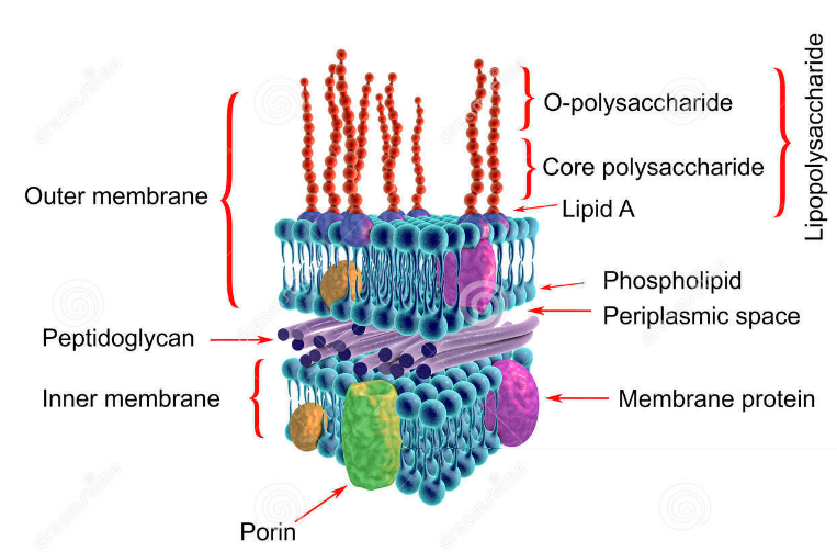
The outer membrane is present in gram negative bacteria that surround the cell wall of bacteria. This membrane is composed of lipoprotein, lipopolysaccharides and phospholipids. The lipopolysaccharide is composed of two portions i.e. polysaccharides and lipid A portion. Lipid A portion of this membrane is an endotoxin in nature. This endotoxin is the distinguishing feature of gram-negative bacteria. The lipid A portion induces fever and shock by lowering the blood pressure of the whole body. Vasodilation of the blood vessel occurs when the whole BP of the body becomes low. This can cause the death of the organism.
Porins:
This membrane has many pores that are called as porins. Porins are permeable lipo-proteins and allow the transportation of substrate and nutrient to move in the cell. These porins can either be selective or non-selective. Selective porins allow the transportation of selective substances while non-selective porins allow any substance to move in the cell. The outer membrane has many structures that help the bacteria to attach to the surfaces.
Periplasmic space:
Periplasmic space is the area between the outer membrane and cell wall that is present in gram negative bacteria. This space plays a vital role in the bacteria as it stores macromolecules. Macromolecules help in the metabolism of the cell and environmental response. It contains many binding proteins that bind amino acids, vitamins, iron and enzymes. These substances are essential for the nutrition of bacteria.
Teichoic acid:
Teichoic acid is present in the gram negative bacteria cell wall. It is present in minute quantity and present as lipo-teichoic acid. These are polymers of polyglycerol that are supplemented with phosphates and some sugar residues. Their function is unknown up till now. Some theories revealed that they are essential for the assembly of peptidoglycans monomers in the cell wall. Examples of gram negative bacteria:
- Escherichia coli
- Salmonella
- Shigella
- Proteus
- Vibrio
- Campylobacter
Staining of gram negative cell wall: The gram negative bacterial cell wall has less component of peptidoglycan. This low content of peptidoglycan doesn’t retain the primary dye colour. Primary stain and mordant bind with the peptidoglycan content of cell wall and make a very weak bonding as CV-PG-I complex. They have a high content of lipid content in the cell wall. This CV-PG-I complex and lipids of the cell wall are washed by using alcohol. Alcohol dissolves the lipid content of the cell wall and thus causes empty spaces between the cell wall. Saffarine enters in the empty spaces of the cell wall and binds with them. The secondary dye retains in the empty space and thus, don’t wash away. Then, during microscopy, these bacteria appear as a pink colour under the microscope.
Resistance mechanism:
They are naturally resistant to Beta-lactams antibiotics. They have a low content of peptidoglycan that’s why they are least effective by beta-lactams antibiotics. But, they have three mechanisms of resistance against penicillin.
Penicillinase enzyme:
Bacteria synthesize the Penicillinase enzymes that degrade the penicillin ring. Penicillin antibiotic activity depends on the beta-lactam ring. When the ring are broken by enzymes, its activity stop and bacteria survive in the environment.
Modified target site:
Antibiotics always work by binding to its target site. Bacteria change its target site for the antibiotics. When the target sites are changed, antibiotics are unable to bind with them. The unbinding of antibiotic with the target site destroys the effect of the antibiotic.
Efflux pump:
Bacteria have some modified proteins that pump the penicillin out from the cell. when penicillin moves out, bacteria survive and able to grow.