
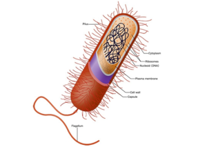
The capsule is the structure that acts as an invisible cloak and helps the bacteria from phagocytosis.
The outermost protective layer (highlighted in light blue). It acts like an “invisibility cloak” by hiding cell antigens from host phagocytes (white blood cells) and preventing the bacterium from being engulfed. It has a sticky, high-water-content nature that enables attachment to host tissues and shields the cell from drying out (desiccation).
Characteristics of capsule:
The characteristics of the capsule are as follows:
- This capsule can be found either on gram positive or gram negative bacteria.
- The capsule is a 0.2µm thick viscous layer.
- The capsule is present outside of the cell wall of bacteria.
- The capsule is different from the cell wall of bacteria.
- It is usually composed of polysaccharide but sometimes it is composed of glycoproteins.
- This capsule can’t be washed by using alcohol or other chemicals.
- This capsule can be stain separately to visualize the bacteria.
- This capsule can be loosely attached or tightly attach to the cell wall.
- Bacteria that have capsule outer to the cell wall is called as encapsulated bacteria.
Composition of capsule:
The capsule composition is different from bacteria to bacteria. The capsule has 2% polysaccharide and 98% water. Mostly, the capsule is composed of polysaccharide. Polysaccharides are the long chains of carbohydrate. The monomeric subunit is combined to form long chains of carbohydrate. The glycosidic linkages are responsible for the binding of different monomeric units. On hydrolysis, they give the monomeric components that constitute the polysaccharide unit. Some bacterial capsule is composed of glycoproteins. Glycoproteins are the structure in which the amino acids of proteins are attached to the carbohydrate group. Carbohydrate groups attach with the chain of polypeptides by covalently bonding. These oligosaccharides attach with the peptide chains during translation and post-translation modification. Some bacteria contain polypeptide D-glutamate capsule. This type of capsule is present in Bacillus antharacis. D-glutamate is the alpha amino acid that is synthesize by many living organism. It is a non-essential amino acid that can be synthesized by the human body. This D-glutamate forms the capsule of bacteria. E.coli capsule has peptidoglycan and muramic acids units in it. They are tightly packed to the capsule and don’t allow the stain to adhere to the capsule.
Function:
The capsule is tightly or loosely bound to the bacteria cell. Capsule performs many functions in the bacterial cell. Some of the functions of the capsule are as follows:
-
Prevent phagocytosis:
The capsule is virulent in nature. It is responsible for the pathogenesis of bacteria. It invades the immune system and allows bacteria to produce disease. It prevents the phagocytosis of the cell. Phagocytosis is the process in the immune regulation of host. In this process, first the pathogen must enter the body and phagocytosis occurs. Phagocytosis is an important process that is performed by phagocytes. Phagocytes phagocytized the pathogens or particles. When a pathogen is engulfed by a phagocyte, an intracellular vesicle called a phagosome captured it. Phagosome fuses with the lysosome to form a phagolysosome. The lysosome contains many digestive enzymes and chemicals that produce respiratory burst. It releases free radicals into the phagolysosome that killed the pathogen. The capsule has many methods to avoid phagocytosis.
- It remains confined to the surfaces where the macrophages are inaccessible.
- It remains in many tissues and surface tissue have less accessibility of macrophages.
- It provokes the inflammation that doesn’t allow the immune system to recognize the pathogen.
- It inhibits the phagocytosis chemotaxis. For example; streptolysin of Streptococcus pneumoniae inhibits the neutrophils. If neutrophils don’t come, no immune response occurs.
- It covers the pathogen by the membranes. This membrane helps the bacteria to invade from the immune system. The immune system doesn’t recognize the bacteria because it identifies it as self-molecule due to the presence of the membrane.
Attachment:
Capsule helps bacteria to attach to the surfaces. The capsule has a sticky nature that attaches with the surface of the host cell. The fruitful attachment is responsible for the development of the disease. Without the attachment, bacteria don’t cause disease. E.g. pneumonia is caused by the capsulated Streptococcus pneumoniae. If the capsule of bacteria removed by using heat or chemical, the non-capsulated bacteria don’t cause disease in the host.
Protection:
The capsule is the polysaccharide layer that protects the bacterial cell from a harsh environment. It protects the cell from mechanical injury, heat and unfavourable environment. Capsule doesn’t allow water to move outside the cell and thus prevent the cell from desiccation. In a warm environment, bacteria water loss and bacteria.
Source of nutrition:
The capsule may provide the source of nutrition for the bacteria. Bacteria may face a shortage of nutrient when growing in the environment. When nutrients deplete, the bacteria use a capsule as a source of carbohydrate. It degrades the capsule and then used it as a source of nutrition.
Types of capsule:
There are two types of the capsule that are present in bacteria. The classification is done by using the thickness of the capsule.
Macro-capsule
This type of capsule has 0.2µm or more thickness. It is a thick membrane that can be observed through a compound microscope.
Microcapsule
The capsule has a thickness of less than 0.2µm. It is a thin membrane that is visible under an Electron microscope only.
Capsule staining:
The capsule can be observed by means of different staining. Staining imparts colour to the bacterial capsule. When colour is imparted to the capsule, the capsule is easily identified under the microscope. There is different staining that is used to observe a capsule.
India ink:
India ink is an acidic stain that has a negative charge. A bacterial cell has a negative charge on its surface. Due to the repulsive forces, India ink can’t penetrate into the bacterial cell. India ink imparts black colour to the background because of attractive forces. The capsule appears as a hollow space in the black background. It is an easy way to identify the bacterial capsule.
Maneval’s capsule stain:
The two stains are used in this method. The one is Congo red that is an acidic stain. It imparts a bluish grey colour. The other stain is also acidic stains that are acid fuchsin. It imparts a pink colour to the bacterium. They both have positive charges. The capsule appears as a clear hollow area between the pink-stained bacterium and the bluish-grey stained background.
Copper sulfate:
Copper sulfate is also used to stain the bacterial capsule.