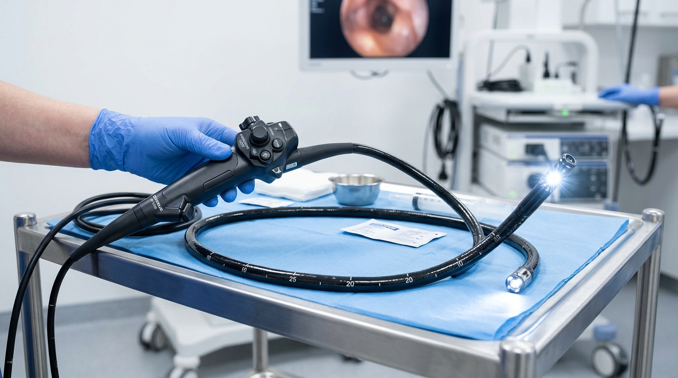
A bronchoscope is a specialized endoscope designed for direct visual examination of the tracheobronchial tree. In clinical microbiology and pulmonary medicine, the instrument is indispensable for inspecting mucosal airways, retrieving transbronchial biopsies, aspirating sterile sputum samples, and obtaining bronchoalveolar lavage (BAL) fluid for microbial isolation and sensitivity testing.
Working Principle of the Bronchoscope
A bronchoscope operates by guiding a lighted, articulating optical tube into the respiratory tract via the oral or nasal cavities, or through a tracheostomy stoma.
Light Source (LED/Xenon) ──> Fiberoptic Bundles ──> Distal Lens ──> CCD/CMOS Sensor ──> Video Processor ──> Digital Monitor- Illumination: Light from an external xenon or solid-state LED source is transmitted through coherent fiberoptic bundles down the insertion cord to illuminate the dark lumen of the bronchi.
- Image Capture:
- In traditional fiberoptic bronchoscopes, objective lenses focus the reflected light onto an imaging bundle that delivers the optical picture directly to an eyepiece.
- In modern video bronchoscopes, a miniature Charge-Coupled Device (CCD) or CMOS chip mounted at the distal tip captures high-definition digital signals and transmits them to an external video processor, projecting real-time images onto high-resolution medical displays.
- Auxiliary Functions: The integrated internal working channel permits the simultaneous delivery of topical anesthetics, supplemental oxygen, suction evacuation of mucus or blood, and the insertion of biopsy forceps, cytology brushes, and aspiration catheters.
Structural Anatomy and Components
A standard flexible video bronchoscope consists of three primary functional zones: the control handle (body), the flexible insertion cord, and the universal connector cable.
1. Control Handle and Body
- Control Lever (Angulation Lever): Ergonomically positioned for thumb control, moving this lever flexes the distal tip up to 180° upward and 130° downward in the vertical plane. Lateral adjustments are accomplished by rotational movements of the operator’s wrist and shoulder.
- Eyepiece / Diopter Ring: Present on fiberoptic units to focus the image for the examiner’s eye. On video bronchoscopes, this is replaced by an ergonomic grip with programmable image capture buttons.
- Suction Valve: A spring-loaded valve depressed by the index finger to apply negative pressure, clearing secretions, blood, and fluids into a collection canister.
- Working Channel Port (Biopsy Port): The entrance through which sterile saline, local anesthetics (e.g., 2% lidocaine), micro-instruments, and cytology brushes are introduced.
2. Flexible Insertion Cord
- Distal Articulation Section: A segmented vertebrae-like metal sheath covered by a flexible waterproof rubber jacket that bends precisely in response to control lever cables.
- Distal Tip: Houses the objective lens, dual illumination light guide lenses, nozzle for flushing, and the exit aperture of the instrument channel.
3. Light and Video Connector
- Links the bronchoscope to the cold light source, video processor, and air/water insufflation pumps.
Flexible vs. Rigid Bronchoscopes
| Feature | Flexible Bronchoscope | Rigid Bronchoscope |
|---|---|---|
| Material | Pliable silicone / polyurethane | Straight, rigid stainless steel tube |
| Anesthesia | Local anesthesia with conscious sedation | General anesthesia with neuromuscular blockade |
| Airway Access | Trachea down to subsegmental bronchi | Mainstem bronchi and trachea only |
| Primary Use | Diagnostic biopsies, BAL, cytology, staging | Foreign body removal, massive hemoptysis, laser debulking |
| Patient Comfort | High; outpatient day procedure | Requires operating theater |
Clinical Indications and Diagnostic Utility
Bronchoscopy is deployed for both diagnostic evaluation and therapeutic interventions.
Diagnostic Applications
- Microbiological Sampling:
- Bronchoalveolar Lavage (BAL): Instilling and aspirating sterile saline aliquots into a lung segment to harvest alveolar microorganisms, crucial for diagnosing Pneumocystis jirovecii, Mycobacterium tuberculosis, cytomegalovirus, and invasive fungal pathogens (Aspergillus).
- Protected Specimen Brushing (PSB): A telescoping catheter with a wax-plug brush used to obtain uncontaminated quantitative bacterial cultures in suspected ventilator-associated pneumonia (VAP).
- Investigation of Unexplained Hemoptysis: Identifying mucosal ulcers, endobronchial malignancies, or vascular malformations.
- Assessment of Persistent Cough and Stridor: Evaluating foreign body aspiration, dynamic airway collapse, or tracheobronchomalacia.
- Staging Lung Carcinoma: Direct biopsy of mucosal lesions and transbronchial needle aspiration (TBNA) of mediastinal lymph nodes under Endobronchial Ultrasound (EBUS) guidance.
Therapeutic Applications
- Removal of aspirated foreign objects (peanuts, dental fragments, small hardware).
- Suction clearance of tenacious mucous plugs in atelectasis.
- Placement of self-expanding silicone or metallic tracheobronchial stents.
- Endobronchial electrocautery, cryotherapy, or laser ablation of obstructive airway tumors.
Contraindications and Pre-Procedure Safety
Absolute and Relative Contraindications
- Severe refractory hypoxemia (inability to maintain PaO₂ > 60 mmHg on supplemental O₂)
- Unstable hemodynamic status or recent myocardial infarction
- Severe untreated coagulopathy or thrombocytopenia (platelets < 50,000/µL)
- High-grade tracheal stenosis threatening complete occlusion
- Severe intractable cardiac arrhythmias
Potential Risks and Complications
- Hypoxemia and Bronchospasm: Reactive airway constriction triggered by mechanical stimulation.
- Hemorrhage: Particularly following transbronchial lung biopsy.
- Pneumothorax: Occurs in 1–3% of transbronchial parenchymal biopsies.
- Nosocomial Infection: High-level disinfection failures can transmit multidrug-resistant pathogens (Pseudomonas aeruginosa, Mycobacterium spp.).
Procedure Protocol
- Pre-Procedural Preparation: Patients fast for at least 6 to 8 hours prior to the procedure. Baseline coagulation profiles (PT/INR, aPTT) and blood counts are reviewed.
- Sedation and Anesthesia: The upper airway is anesthetized using topical 2% to 4% lidocaine spray. Conscious sedation with intravenous midazolam and fentanyl is administered under continuous pulse oximetry and ECG monitoring.
- Airway Navigation: The bronchoscope is introduced transnasally or transorally through a bite block, traversing the vocal cords during inspiration.
- Inspection and Sampling: The tracheobronchial anatomy is surveyed methodically from the vocal cords to segmental sub-branches. Diagnostic washes, brushings, or biopsies are collected into sterile containers and immediately transported to the microbiology and pathology laboratories.