Introduction
Agarose gel electrophoresis is a standard tool in clinical and molecular microbiology for verifying 16S rRNA PCR amplicons (~1,500 bp), screening multiplex antimicrobial resistance panels (such as blaKPC, blaNDM, blaOXA-48, and mecA), and checking plasmid preparations.
When bands appear smeared, faint, or crooked, troubleshooting starts with a simple diagnostic check: compare the sample lanes against the molecular weight ladder and positive PCR control. If both the ladder and sample lanes fail, the defect lies in gel preparation, running buffer exhaustion, or electrical settings. If the ladder resolves cleanly while sample lanes fail, the problem originates upstream during bacterial culture harvesting, cell lysis, or DNA purification.
Pre-Analytical Microbiology Factors
Many apparent gel failures occur before samples ever enter the wells.
- Incomplete Gram-Positive Lysis: Thick peptidoglycan walls in Staphylococcus aureus resist standard lysozyme because peptidoglycan O-acetyltransferase (OatA) modifies muramic acid residues. Supplementing lysis buffers with lysostaphin (10 to 200 µg/mL) for staphylococci or mutanolysin (100 to 250 U/mL) for streptococci and enterococci prevents weak or false-negative PCR results.
- Nuclease Carryover and Shearing: Harvesting cultures past exponential growth (ideal OD600 is 0.6 to 0.8 for plasmids or 0.8 to 1.0 for genomic DNA) triggers endogenous autolysins that fragment DNA. Using EndA+ Escherichia coli strains (such as HB101) introduces Endonuclease I carryover that degrades plasmids into smears; switch to EndA- strains (such as DH5α or TOP10), include Proteinase K, and invert lysates gently rather than vortexing during alkaline lysis.
- Plasmid Conformation Artifacts: Uncut bacterial plasmids naturally separate into multiple bands: compact supercoiled monomers (Form I) migrate fastest, linear monomers (Form III) migrate at true size, and relaxed open circles (Form II) migrate slowest. Always run a single-cutter restriction-linearized control alongside uncut plasmid preparations.
- PCR Inhibitors and Salt Carryover: Co-purified exopolysaccharides, teichoic acids, and chaotropic salts depress purity ratios (A260/A230 < 1.5) and inhibit Taq polymerase. Diluting templates 1:10 or 1:100 or adding 0.4 µg/µL BSA overcomes inhibition.
Fixing Smeared Bands
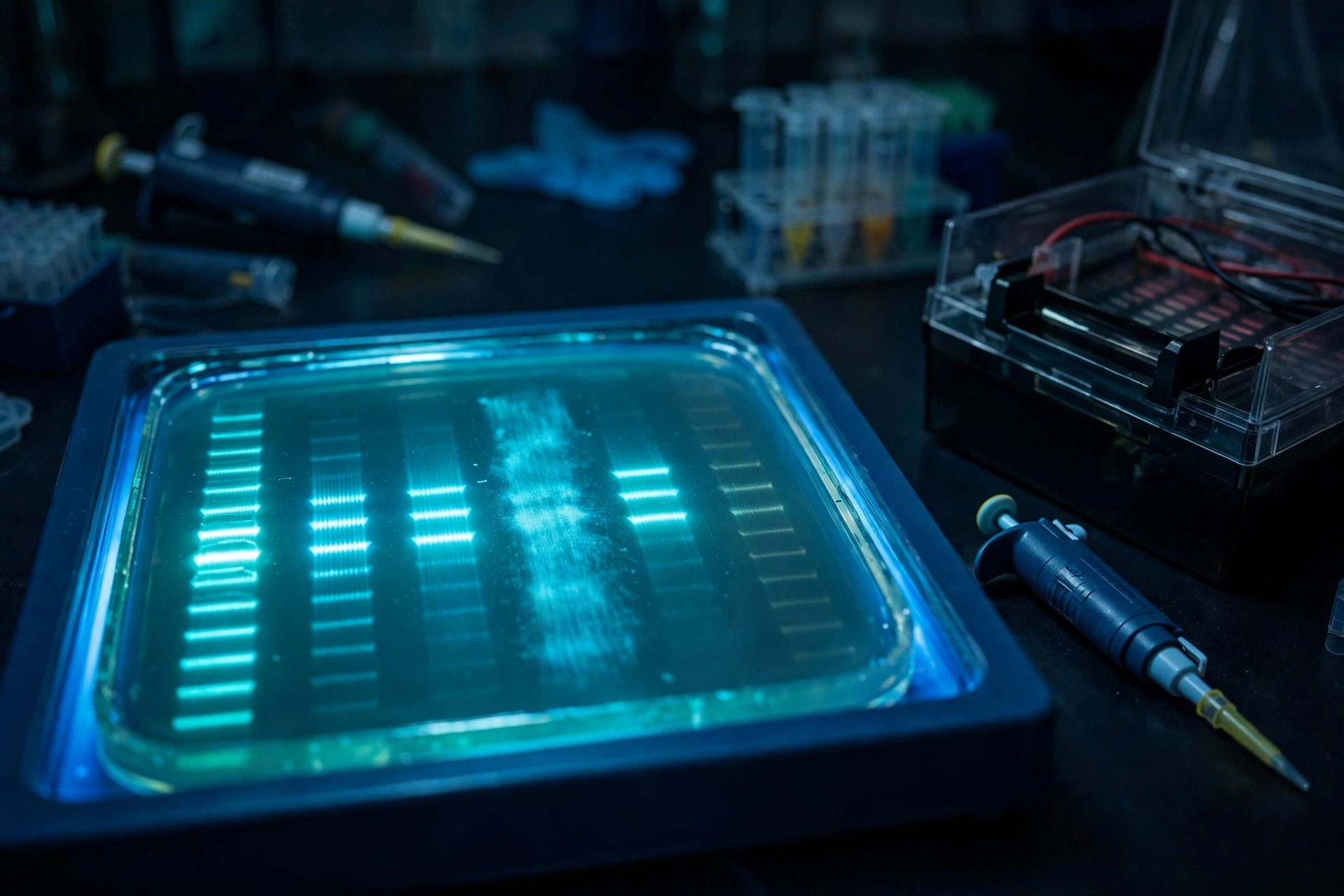
Smeared bands appear as continuous vertical streaks or fuzzy halos across molecular weight ranges.
- DNA Degradation or Mechanical Shearing: Biological fragmentation from EndA+ nucleases, stationary-phase autolysis, or repeated freeze-thaw cycles produces smears extending downward from the wells. Handle high-molecular-weight genomic DNA with wide-bore tips and store extracts in EDTA-containing buffer.
- Sample Overloading: Exceeding 0.1 to 0.2 µg (100 to 200 ng) of DNA per millimeter of well width clogs gel pores and creates trailing comet smears. Load 50 to 200 ng per single sharp PCR band (or ≤100 ng per band in precast GelRed gels).
- Residual RNA or Bound Enzymes: Omitting RNase A (100 µg/mL) in plasmid minipreps leaves low-molecular-weight RNA smears, while bound restriction enzymes or ligases retard DNA mobility. Column-purify enzymatic digests or add SDS loading dye with brief heating prior to loading.
- Gel Casting and Buffer Defects: Gels thicker than 5 mm trap heat and blur bands. Cast 3 to 4 mm thick slabs, keep the comb slightly elevated above the tray floor so wells do not leak underneath, and image gels immediately after turning off the power.
Correcting Faint and Missing Bands
Faint or absent bands result from low nucleic acid yield, dye depletion, or electrophoresis setup errors.
- Mismatched Gel Concentration and Tracking Dye Masking: Use 0.7% to 1.0% agarose for large plasmids and 16S rRNA products (~1,500 bp), and 2.0% to 3.0% agarose for short multiplex resistance amplicons (100 to 500 bp). Avoid bromophenol blue when visualizing ~200 to 370 bp fragments on 1% gels, as the dye front casts an optical shadow over co-migrating DNA; switch to orange G (~50 bp) instead.
- Cathodic Ethidium Bromide (EtBr) Migration: Positively charged EtBr migrates toward the negative cathode, opposite to DNA, stripping stain from the lower portion of the gel during long runs. Supplement the running buffer with 0.5 µg/mL EtBr or post-stain the gel after electrophoresis.
- Chaotropic Salt Denaturation: Residual guanidine salts from spin columns can denature double-stranded amplicons into faint, fast-migrating single-stranded bands. Add 10 mM NaCl, heat at 95°C for 1 minute, and cool slowly to room temperature to reanneal the DNA.
- Reversed Polarity or Over-Running: Always verify that sample wells sit at the black cathode (“run to red”) and stop electrophoresis when the leading dye reaches 75% to 80% of the gel length.
Eliminating Crooked, Smiling, and Frowning Bands
Geometric distortion reflects uneven heat dissipation, buffer exhaustion, or physical casting errors.
- Gel-Wide Smiling and Frowning: Running above 5 to 10 V/cm generates central Joule heating. Because the center of the gel stays warmer than the buffer-cooled edges, central lanes migrate faster and form a U-shaped smile, whereas severe central overheating or edge field concentration creates an inverted frown. Calculate voltage strictly using inter-electrode distance (keep routine runs at ≤5 V/cm), maintain a 2 to 5 mm buffer overlay, and load 1× loading buffer into unused outer lanes.
- Buffer Exhaustion: 1× TAE (40 mM Tris, 20 mM acetate, 1 mM EDTA) offers superior downstream enzymatic recovery for cloning and sequencing but acidifies after 2 to 3 hours. Switch to 1× or 0.5× TBE (89 mM Tris, 89 mM borate, 2 mM EDTA) for runs exceeding 2 hours or for sharp separation of fragments under 1,000 bp. Always cast the gel and fill the tank from the same fresh 1× buffer batch.
- Intra-Lane Skewing and Slanted Bands: High sample salt (>50 to 100 mM) diffuses faster at lane edges than in the lane core, warping individual bands into local U-shapes. Desalt samples via ethanol precipitation or spin columns, or start electrophoresis slowly at 15 to 30 V for 10 minutes before increasing voltage. Cool molten agarose to 55 to 60°C before pouring onto a level surface so acrylic trays do not warp.
Quick Troubleshooting Comparison
| Band Phenotype | Primary Causes | Immediate Corrective Action |
|---|---|---|
| Vertical smear from well or >10 kb | EndA+ nuclease carryover, stationary-phase autolysis, or vortex shearing | Use EndA- host (DH5α), harvest at OD600 0.6 to 0.8, invert gently, and include Proteinase K |
| Bright band with trailing comet tail | Sample overload (>0.1 to 0.2 µg/mm well width) | Dilute sample to 50 to 200 ng per sharp band (≤100 ng in precast GelRed) |
| Sample faint/blank; ladder crisp | Incomplete Gram-positive lysis or PCR inhibition | Add lysostaphin or mutanolysin; dilute template 1:10/1:100 or add 0.4 µg/µL BSA |
| Upper ladder bright; lower bands faint | Cationic EtBr migrating backward toward cathode | Add 0.5 µg/mL EtBr to running buffer or post-stain gel for 15 to 30 minutes |
| Gel-wide smiling or frowning arc | High voltage (>5 V/cm), exhausted buffer, or shallow overlay | Lower voltage to ≤5 V/cm (electrode-to-electrode), use fresh 1× buffer, and cover gel by 2 to 5 mm |
| Single-lane U-shape or crooked slant | High sample salt, unlevel casting tray, or slanted comb | Desalt sample, start run at 15 to 30 V briefly, level casting tray, and align comb vertically |
Visualization, Downstream Cloning, and Biosafety
Selecting the right stain and light source protects both recombinant cloning efficiency and laboratory safety.
- Post-Staining for Sizing Accuracy: Precasting EtBr slows migration by roughly 10% and unwinds supercoiled plasmids, while precasting GelRed above 100 ng per band can shift or smear bands. Post-stain unstained gels when exact sizing or plasmid topology analysis is required.
- Blue-Light LED vs. UV Transillumination: Short-wave 254 nm UVC induces thymine dimers and strand breaks within seconds, while 302 to 312 nm UVB still risks DNA nicking during band excision. Pairing SYBR Safe or GelGreen with a 470 nm blue-light LED transilluminator preserves bacterial transformation efficiency and eliminates UV eye and skin hazards.
- EtBr Waste Decontamination: Never use sodium hypochlorite (bleach) to deactivate EtBr, as incomplete oxidation leaves mutagenic byproducts, and note that autoclaving does not destroy EtBr. Deactivate liquid EtBr buffers using activated charcoal filtration or commercial adsorbent bags, verify zero fluorescence under handheld UV light, and route all EtBr-contaminated gels, wipes, and adsorbents for hazardous waste incineration.