- 1 Microbes and the Vaginal Mucosa
- 2 The Vaginal Microbiome
- 3 Pathogenesis of Bacterial Vaginosis
- 4 Historical Perspective
- 5 Taxonomy and Classification
- 6 Microscopic and Phenotypic Characteristics
- 7 Growth Requirements and Culture
- 8 Diseases and Clinical Pathology
- 9 Signs and Symptoms
- 10 Diagnostic Procedures
- 11 Antimicrobial Treatment Strategies
- 12 Risk Factors and Prevention
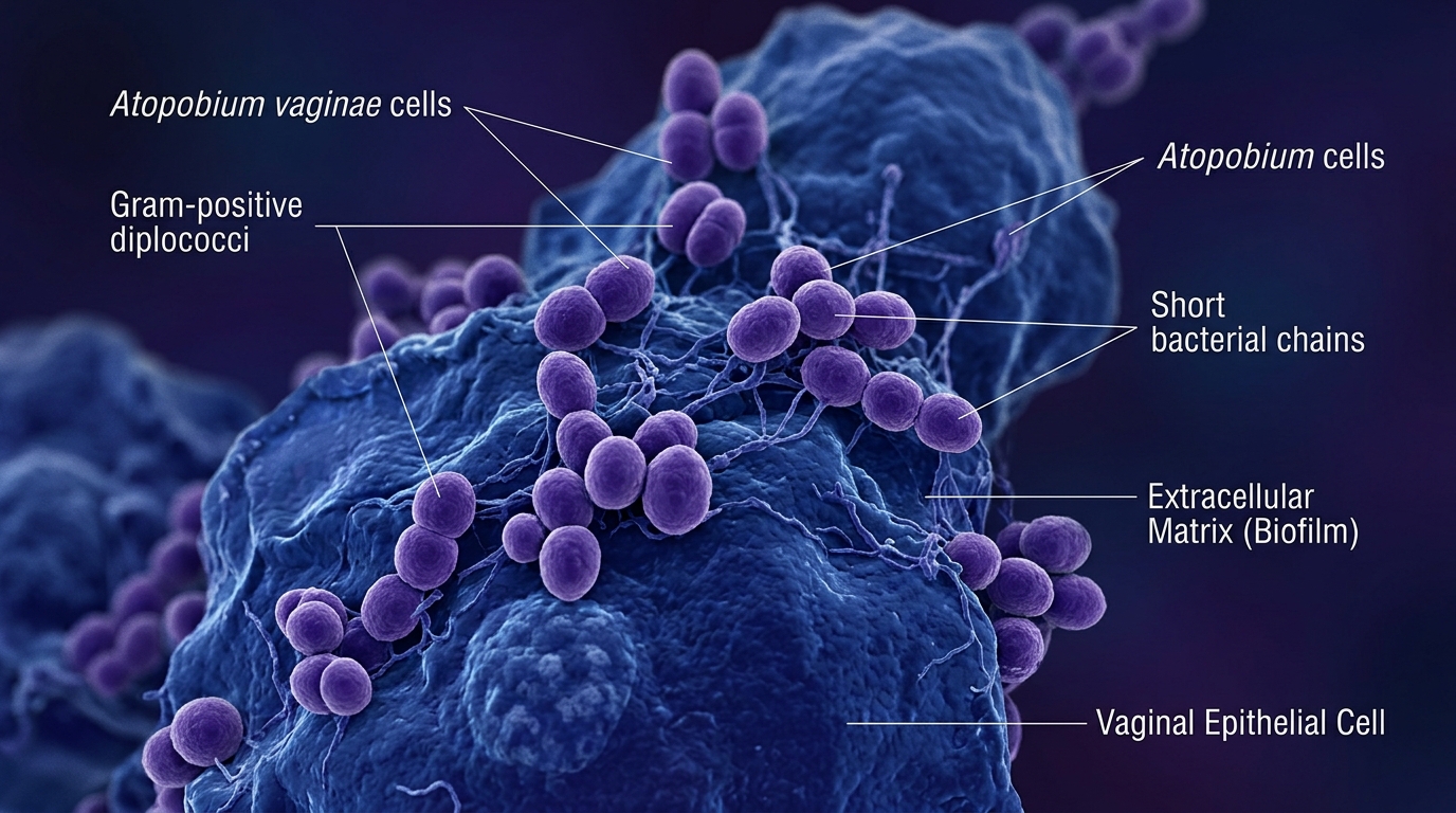
Atopobium vaginae is a fastidious, anaerobic Gram-positive coccobacillus that plays a critical pathogenic role in recurrent and refractory bacterial vaginosis (BV). While historically overlooked due to culture difficulty, advanced molecular assays have demonstrated that A. vaginae acts synergistically with Gardnerella vaginalis to produce robust polymicrobial biofilms on the vaginal epithelium.
Microbes and the Vaginal Mucosa
The healthy female reproductive tract maintains a mutualistic relationship between the vaginal mucosal epithelium and resident microflora. In healthy reproductive-age women, this microbiome is dominated by Lactobacillus species, which ferment glycogen-derived carbohydrates into lactic acid, maintaining an acidic vaginal pH between 3.5 and 4.5.
Vaginal Glycogen ──(Lactobacillus spp.)──> Lactic Acid + H₂O₂ ──> Acidic pH (3.5–4.5) ──> Pathogen InhibitionWhen this protective equilibrium is disrupted, lactobacilli counts drop dramatically, creating a permissive ecological niche for diverse anaerobic and facultative organisms, culminating in the syndrome known as bacterial vaginosis.
The Vaginal Microbiome
The vaginal microbiota is typically categorized into Community State Types (CSTs) based on dominant lactobacilli:
- Lactobacillus iners: Highly dynamic and frequently observed in transitional states between eubiosis and dysbiosis.
- Lactobacillus crispatus: Strongly associated with stability, low pH, and high hydrogen peroxide (H₂O₂) production.
- Lactobacillus gasseri and Lactobacillus jensenii: Common protective acid producers.
The normal microbial load of lactobacilli prevents the colonization and ascent of urogenital pathogens such as Neisseria gonorrhoeae, Chlamydia trachomatis, and HIV. In addition to lactic acid, lactobacilli produce bacteriocins and hydrogen peroxide that suppress competing anaerobes.
Pathogenesis of Bacterial Vaginosis
Bacterial vaginosis is characterized by a dysbiotic shift from a homogeneous lactobacilli population to a dense, complex polymicrobial community of anaerobic organisms, including:
- Gardnerella vaginalis
- Atopobium vaginae
- Mobiluncus curtisii and M. mulieris
- Prevotella bivia and Prevotella disiens
- Megasphaera and Clostridial-like species (BVAB1–3)
Along with Gardnerella, A. vaginae forms structured, adherent biofilms on the vaginal epithelium that protect the constituent bacteria against both host immunological clearing mechanisms and conventional antibiotic penetration.
Historical Perspective
Atopobium vaginae was first isolated and described in Sweden in 1999 by Rodriguez et al. from the vaginal flora of a healthy woman. Members of the genus Atopobium had previously been isolated from human abdominal wounds, fecal matter, and dental abscesses. In 2003, its medical significance was definitively established after it was recovered in pure culture from a patient suffering from a tubo-ovarian abscess, highlighting its potential for invasive pelvic disease.
Taxonomy and Classification
Atopobium vaginae belongs to the phylum Actinobacteria (now frequently classified as Fannyhessea vaginae).
| Classification Rank | Taxonomic Name |
|---|---|
| Domain | Bacteria |
| Phylum | Actinobacteria |
| Class | Coriobacteriia |
| Order | Coriobacteriales |
| Family | Atopobiaceae |
| Genus | Atopobium (Fannyhessea) |
| Species | Atopobium vaginae |
| Binomial Name | Atopobium vaginae (Rodriguez et al., 1999) |
Microscopic and Phenotypic Characteristics
Under Gram stain examination, Atopobium vaginae presents as small Gram-positive elliptical cocci or short coccobacilli measuring 0.6–0.9 µm. They appear singly, in pairs, or in short chains. The organism is:
- Non-motile
- Non-spore-forming
- Strictly anaerobic
- Acid phosphatase positive
Growth Requirements and Culture
Atopobium vaginae is notoriously fastidious. It requires enriched culture media such as Columbia blood agar supplemented with 5% sheep or human blood, incubated in an anaerobic chamber (80% N₂, 10% CO₂, 10% H₂) at 37°C for 48 to 72 hours. Colonies are minute, pin-point, translucent to grayish, and non-hemolytic.
Diseases and Clinical Pathology
The primary clinical condition caused by A. vaginae is bacterial vaginosis, but it is also implicated in:
- Pelvic Inflammatory Disease (PID): Ascent into the endometrium and fallopian tubes.
- Late-trimester Miscarriage and Preterm Birth: Secondary to chorioamnionitis.
- Endometritis and Post-gynecological Infection: Surgical site infections following hysterectomy or cesarean section.
Biofilm Mechanism
In BV pathogenesis, lactobacilli depletion results in elevated vaginal pH. Gardnerella vaginalis first adheres to vaginal epithelial cells, creating an initial scaffold. Atopobium vaginae then integrates into this matrix, producing dense, recalcitrant biofilms. Microbial metabolism shifts from lactic acid production to short-chain fatty acids (succinate, butyrate, acetate) and volatile polyamines (putrescine, cadaverine), which generate the characteristic fishy amine odor.
Signs and Symptoms
While some women remain asymptomatic, clinical BV typically manifests as:
- Elevated Vaginal pH: Exceeding 4.5.
- Discharge: Thin, homogeneous, grayish-white vaginal discharge coating the mucosal walls.
- Odor: Distinctive amine “fishy” odor, notably intensified after unprotected intercourse.
- Vulvovaginal Irritation: Mild pruritus, burning sensation, and dyspareunia.
Diagnostic Procedures
1. Amsel Clinical Criteria
A clinical diagnosis of bacterial vaginosis requires at least 3 of the following 4 criteria:
- Thin, homogeneous, grayish-white discharge.
- Vaginal secretion pH > 4.5.
- Positive Whiff test (release of fishy amine odor upon adding 10% KOH to secretions).
- Presence of “clue cells” (vaginal epithelial cells studded with adherent bacteria, obscuring border margins) on wet mount microscopy (> 20% of epithelial cells).
2. Nugent Score (Gram Stain)
Microscopic scoring of vaginal smears evaluating the relative proportions of large Gram-positive rods (Lactobacillus morphotypes), small Gram-variable rods (Gardnerella/Bacteroides morphotypes), and curved Gram-variable rods (Mobiluncus morphotypes). A score of 7 to 10 is diagnostic for BV.
3. Quantitative Real-Time PCR (qPCR)
Because culture is technically challenging and slow, quantitative multiplex PCR assays targeting the 16S rRNA gene of Atopobium vaginae serve as sensitive, specific diagnostic tools. Detecting high bacterial loads of A. vaginae correlates strongly with recurrent and treatment-refractory BV.
Antimicrobial Treatment Strategies
The pharmacological management of Atopobium vaginae requires special attention because many strains exhibit intrinsic or high-level acquired resistance to metronidazole:
- Metronidazole Limitations: While metronidazole (500 mg oral twice daily for 7 days or 0.75% vaginal gel) is the first-line standard for general BV, A. vaginae frequently possesses high minimum inhibitory concentrations (MICs > 128 µg/mL). Patients treated with metronidazole alone often experience rapid relapse because the biofilm-embedded A. vaginae survives.
- Clindamycin: Clindamycin (300 mg oral twice daily for 7 days or 2% vaginal cream at bedtime for 7 days) demonstrates superior in vitro and in vivo activity against A. vaginae and represents the preferred therapy for metronidazole-refractory cases.
- Biofilm Disruptors: Emerging adjuvant therapies including boric acid and topical dequalinium chloride are being deployed to disrupt the matrix before antibiotic administration.
Risk Factors and Prevention
Risk factors for acquiring BV and A. vaginae colonization include:
- Frequent vaginal douching (disrupts natural lactobacilli flora)
- Multiple sexual partners or recent partner changes
- Cigarette smoking and chronic psychosocial stress
- Lack of barrier contraception
Preventive recommendations focus on avoiding scented feminine hygiene washes, eliminating douching, utilizing barrier contraception, and considering oral or vaginal Lactobacillus probiotics following antimicrobial treatment to promote eubiotic re-colonization.