Mycobacterium abscessus is a rapidly growing, multidrug-resistant non-tuberculous mycobacterium (NTM) found ubiquitously in soil, water, and biofilm environments. Historically viewed as an occasional wound contaminant, it has emerged as a formidable human pathogen responsible for chronic, debilitating pulmonary infections in patients with underlying lung diseases (such as cystic fibrosis and bronchiectasis) and severe skin and soft-tissue infections following surgical or cosmetic procedures.
History and Taxonomy
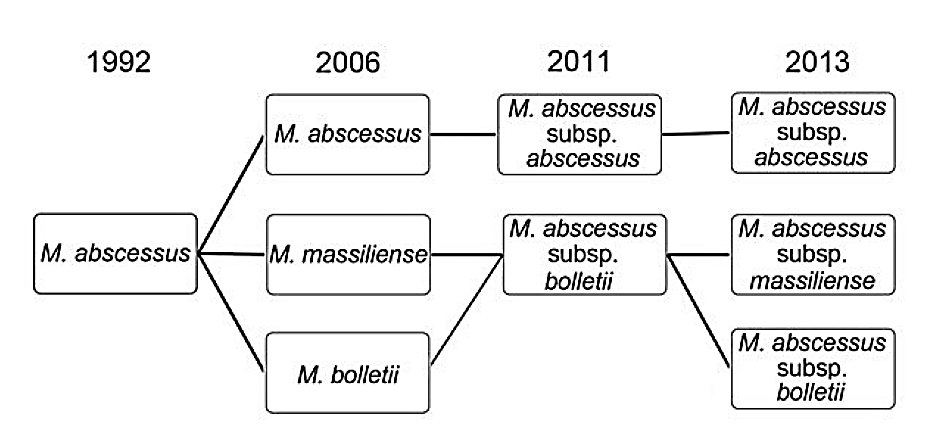
Mycobacterium abscessus was first isolated in 1952 by Moore and Frerichs from a 63-year-old patient presenting with a chronic abscess-like knee infection following trauma. In 1992, Kusunoki and Ezaki distinguished it as a unique species separate from Mycobacterium chelonae.
Modern whole-genome sequencing defines the Mycobacterium abscessus complex (MABC) into three distinct subspecies:
- Mycobacterium abscessus subsp. abscessus
- Mycobacterium abscessus subsp. massiliense
- Mycobacterium abscessus subsp. bolletii
| Classification Rank | Taxonomic Name |
|---|---|
| Domain | Bacteria |
| Phylum | Actinobacteria |
| Class | Actinomycetia |
| Order | Mycobacteriales |
| Family | Mycobacteriaceae |
| Genus | Mycobacterium |
| Species | Mycobacterium abscessus |
| Binomial Name | Mycobacterium abscessus (Moore & Frerichs, 1953) |
Morphology and Physiology
Mycobacterium abscessus is an aerobic, non-motile, non-spore-forming rod measuring 1.0–2.5 µm in length and 0.5 µm in width.
Key microbiological characteristics include:
- Acid-Fast Staining: Strongly acid-fast positive on Ziehl-Neelsen and Kinyoun staining due to its thick, waxy, lipid-rich mycolic acid cell wall.
- Rapid Growth Rate: Classified in Runyon Group IV (Rapid Growers); produces visible colonies on solid media within 3 to 7 days, contrasting with slow-growing mycobacteria like M. tuberculosis (which take 3 to 6 weeks).
- Colony Morphotypes: Exhibits two distinct colony phenotypes:
- Smooth (S) Morphotype: Expresses surface glycopeptidolipids (GPL); common in environmental reservoirs and early asymptomatic colonization.
- Rough (R) Morphotype: Lacks surface GPLs, forms prominent serpentine cords, triggers invasive tissue necrosis, and is associated with chronic, aggressive clinical disease.
- Biochemical Features: Tolerates 5% NaCl and growth on MacConkey agar without crystal violet; negative for nitrate reduction and niacin accumulation.
Transmission and Epidemiology
Mycobacterium abscessus is primarily acquired from environmental reservoirs:
- Medical and Surgical Inoculation: Contaminated medical instruments, unsterilized tap water used for rinsing equipment, and contaminated injection needles during cosmetic surgery (liposuction, mesotherapy), acupuncture, and tattooing.
- Respiratory Acquisition: Inhalation of environmental aerosols. While traditionally considered purely environmental, genomic epidemiological studies have demonstrated probable indirect person-to-person transmission among patients with cystic fibrosis via contaminated hospital surfaces and fomites.
Clinical Manifestations
1. Chronic Pulmonary Disease
The most severe manifestation of MABC infection, occurring almost exclusively in patients with pre-existing lung architecture distortion—especially cystic fibrosis (CF), non-CF bronchiectasis, and prior tuberculosis. Symptoms include:
- Chronic productive cough with purulent sputum
- Hemoptysis (coughing up blood)
- Exertional dyspnea and wheezing
- Malaise, anorexia, low-grade fevers, and cachexia
2. Skin and Soft Tissue Infections (SSTI)
Inoculation following trauma, surgical incisions (e.g., post-median sternotomy), or cosmetic injections produces painful, erythematous, violaceous subcutaneous nodules, boils, draining sinus tracts, and chronic non-healing ulcers.
3. Disseminated Disease
Occurs in profoundly immunocompromised hosts (solid-organ transplant recipients, patients on anti-TNF biologics), causing multifocal osteomyelitis, splenic abscesses, and bacteremia.
Antimicrobial Resistance Mechanisms
Mycobacterium abscessus is notorious as one of the most antibiotic-resistant bacterial species known, rendering standard anti-tuberculosis medications (isoniazid, rifampin, ethambutol, pyrazinamide) completely ineffective:
MABC Resistance ──> Impermeable Mycolic Acid Wall + Efflux Pumps + erm(41) Inducible Macrolide Resistance- Impermeable Cell Envelope: Porins and mycolic acid architecture limit passive drug entry.
- Inducible Macrolide Resistance (erm(41) Gene): Subspecies abscessus and bolletii harbor a functional erm(41) gene encoding an rRNA methyltransferase. When exposed to macrolides (clarithromycin, azithromycin), the enzyme methylates the 23S rRNA binding site, conferring high-level resistance after 14 days of incubation.
- Subspecies Distinction: Subspecies massiliense carries a truncated, non-functional erm(41) gene, remaining stably susceptible to macrolides and responding significantly better to treatment.
Diagnostic Protocol
- Smear Microscopy: Fluorescent auramine-rhodamine and Ziehl-Neelsen staining of sputum or biopsied tissue.
- Culture: Plated onto selective Lowenstein-Jensen, Middlebrook 7H10/7H11 agar, and automated liquid culture systems (BACTEC MGIT).
- Molecular Differentiation: Sanger sequencing or line-probe hybridization of hsp65, rpoB, and the erm(41) locus to confirm species identity and establish inducible macrolide resistance status.
Therapeutic Regimens
Curing M. abscessus requires prolonged multidrug combination therapy spanning 12 to 24 months, managed in consultation with infectious disease specialists:
Intensive Phase (3 to 12 Weeks)
- Intravenous Amikacin (10–15 mg/kg daily)
- Intravenous Cefoxitin (up to 12 g daily in divided doses) or Imipenem
- Intravenous or Oral Tigecycline
- Plus an oral macrolide (Azithromycin 250–500 mg daily), provided erm(41) is non-functional
Continuation Phase (Oral / Inhaled for > 12 Months Post-Culture Conversion)
- Daily Azithromycin
- Inhaled Liposomal Amikacin
- Plus two or three oral agents based on susceptibility: Clofazimine, Linezolid, or Bedaquiline
Adjunctive surgical resection of localized cavitary lesions is frequently required to achieve definitive cure.