- 1 1. Cell Culture: Testing for Replicative Competence
- 2 2. Plaque Assay: Quantifying Infectious Viral Units
- 3 3. RT-PCR & RT-qPCR: Molecular Detection of Genetic Material
- 4 4. Comprehensive Comparison: What Does Each Method Actually Measure?
- 5 5. The Clinical Dilemma: Why RT-PCR Stays Positive While Culture Is Negative
- 6 6. Which Method Is the True “Gold Standard”?
- 7 7. How Modern Virology Laboratories Integrate All Three Modalities
- 8 Conclusion
Imagine receiving a clinical nasopharyngeal swab in a diagnostic virology laboratory. You extract total RNA, run a real-time RT-PCR assay, and observe an amplification curve crossing the baseline at a low cycle threshold (Ct = 18). The sample is unequivocally positive for viral nucleic acid.
Does this result prove the patient is actively shedding infectious, replication-competent virions capable of transmitting disease? Not necessarily.
Now imagine inoculating an aliquot of that same clinical specimen onto a confluent monolayer of susceptible cells. Over the next 48 to 72 hours, you observe progressive cell rounding, syncytium formation, and detachment—classic cytopathic effects (CPE) indicating active intracellular viral replication. Finally, you prepare a serial dilution series, perform a plaque assay with an agarose overlay, and count 45 distinct clear plaques, quantifying an exact infectious viral titer in plaque-forming units per milliliter (PFU/mL).
Each of these three laboratory techniques analyzed the identical biological sample, yet each measured a fundamentally different property of the virus:
The Three Methodological Lenses of Virology:
├── 1. RT-PCR: Molecular Lens ──────> Detects Viral Genetic Material (RNA)
├── 2. Cell Culture: Phenotypic Lens > Demonstrates Replicative Competence in Host Cells
└── 3. Plaque Assay: Functional Lens > Quantifies Discrete Infectious Viral Particles
Which of these diagnostic modalities represents the true “gold standard”? The answer depends entirely on the biological or clinical question being asked. In this guide, we will examine the principles, mechanics, advantages, and limitations of cell culture, plaque assays, and RT-PCR, and explore how modern virology laboratories integrate all three to achieve diagnostic precision.
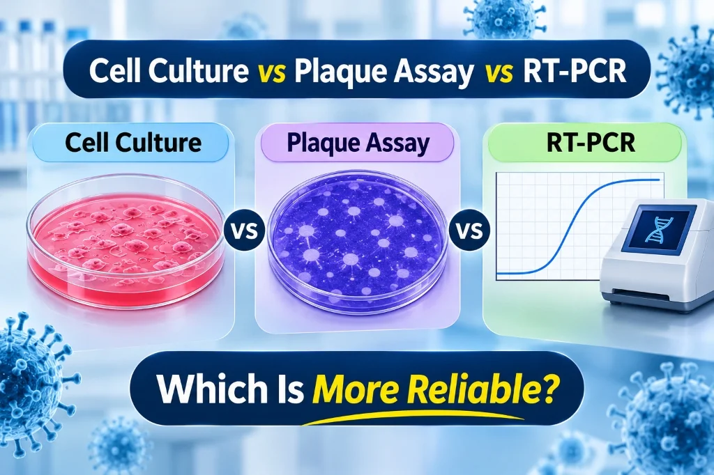
Figure 1: Direct comparison of the three primary virological modalities: cell culture isolation, plaque assay infectivity quantification, and RT-PCR molecular detection.
1. Cell Culture: Testing for Replicative Competence
For nearly a century—originating from the breakthrough work of John Enders, Thomas Weller, and Frederick Robbins in cultivating poliovirus in non-neural tissue—virus isolation in cell culture served as the historical bedrock of diagnostic virology.
The underlying principle is biologically elegant: because viruses are obligate intracellular parasites unable to replicate outside a living host cell, providing an optimized in vitro cellular monolayer determines whether an infectious, replication-competent pathogen is present.
Virus Isolation Workflow:
Clinical Specimen ──> Pre-treatment & Inoculation ──> Incubation (35–37°C) ──> Daily CPE Monitoring
Host Cell Tropism and In Vitro Systems
Different virus families exhibit specific cellular tropisms requiring tailored cell substrates:
- Vero Cells (African Green Monkey Kidney): Widely utilized for coronaviruses (SARS-CoV-2), flaviviruses (Dengue, Zika), and filoviruses.
- MDCK Cells (Madin-Darby Canine Kidney): The standard host system for influenza A and B virus propagation.
- HeLa & A549 Cells (Human Epithelial): Standard lines for adenoviruses, enteroviruses, and respiratory syncytial virus (RSV).
- Primary / Differentiated Air-Liquid Interface (ALI) Cultures: Human bronchial epithelial cells grown at an air-liquid interface to mimic human airway physiology for fastidious respiratory isolates.
Cytopathic Effects (CPE)
When a virus infects a host monolayer, viral shut-off of host macromolecular synthesis and cytotoxic viral proteins induce distinct morphological changes known as cytopathic effects (CPE):
- Cell Rounding & Detachment: Disruption of the cytoskeleton and focal adhesion complexes (common in picornaviruses and enteroviruses).
- Syncytium Formation (Multinucleated Giant Cells): Viral fusion proteins on cell membranes drive adjacent cells to fuse into large multinucleated syncytia (classic hallmark of Respiratory Syncytial Virus, Measles, and Herpesviruses).
- Cellular Vacuolation & Foaming: Accumulation of lipid droplets and swollen organelles (typical of retroviruses and certain arboviruses).
- Complete Monolayer Lysis: Rapid burst destruction of the cell sheet (typical of lytic enteroviruses).
Strengths and Critical Limitations of Cell Culture
- Advantages: Unambiguously demonstrates the presence of viable, replicating virus; provides live physical viral isolates for whole-genome sequencing, antigenic profiling, and antiviral drug screening.
- Limitations: Highly laborious with prolonged turnaround times (3 to 21 days); requires specialized BSL-2, BSL-3, or BSL-4 biocontainment infrastructure; vulnerable to bacterial or fungal contamination; and completely blind to “non-culturable” viruses (e.g., Norovirus, Hepatitis B, and Hepatitis C, which grow poorly or not at all in routine immortalized cell lines).
2. Plaque Assay: Quantifying Infectious Viral Units
If cell culture provides a qualitative answer (“can the virus replicate?”), the plaque assay provides a quantitative biological endpoint: how many infectious virions are present in the sample?
Developed for animal virology in 1952 by Renato Dulbecco (adapting bacteriophage techniques), the plaque assay remains the definitive reference standard for quantifying infectious viral titers.
Plaque Assay Mechanism:
1. Confluent Monolayer ──> 2. Serial 10-Fold Dilutions ──> 3. Semisolid Overlay (Agarose)
4. Localized Viral Foci ──> 5. Fixation & Crystal Violet Staining ──> 6. Clear Plaque Enumeration
Assay Mechanics
- Monolayer Infection: Susceptible host cells are grown to 95–100% confluency in multi-well plates.
- Serial Dilution Inoculation: The viral sample is serially diluted across a 10-fold dilution series (10−1 to 10−7), and a fixed volume (e.g., 0.1 mL or 0.2 mL) is applied to each well.
- Semisolid Viscous Overlay: After an initial absorption period (1 hour), the liquid inoculum is replaced with a semisolid overlay (typically 0.6%–1.0% agarose, methylcellulose, or carboxymethylcellulose). This overlay restricts free convection and diffusion of progeny virions through the fluid phase.
- Radial Plaque Formation: As an initially infected cell lyses, released progeny virions can only infect immediately adjacent neighboring cells. Over multiple replication cycles, this localized chain of infection carves out a microscopic circular zone of dead, detached cells—a plaque.
- Fixation and Staining: The overlay is removed, cells are fixed with formaldehyde or methanol, and the monolayer is stained with crystal violet or neutral red. Living host cells absorb the dye, appearing deep purple, while areas of viral cell destruction remain completely clear.
Calculating Plaque-Forming Units (PFU/mL)
The concentration of infectious virus is calculated using the formula:
PFU/mL = (Number of Plaques × Dilution Factor) / Inoculated Volume (mL)
Worked Laboratory Example: Suppose an analyst counts 45 plaques in a well inoculated with 0.1 mL of a 10−5 viral dilution:
PFU/mL = (45 plaques × 105) / 0.1 mL = 4.5 × 10⁷ PFU/mL
Limitations of the Plaque Assay
- Non-Plaquing Viruses: Many clinically critical viruses (such as Hepatitis C virus and non-cytopathic strains of Influenza) do not lyse host cells sufficiently to form clear optical plaques, necessitating alternative titration methods such as the Median Tissue Culture Infectious Dose (TCID50) or focus-forming assays (FFA).
- Physical vs. Infectious Particle Ratio: A plaque-forming unit represents a single infectious focus, but it does not represent a single physical virion. Due to defective interfering particles, non-functional capsids, or packaging errors, the particle-to-PFU ratio for animal viruses typically ranges from 10:1 to over 10,000:1.
3. RT-PCR & RT-qPCR: Molecular Detection of Genetic Material
Reverse transcription polymerase chain reaction (RT-PCR) revolutionized diagnostic microbiology by decoupling viral detection from the biological requirement of cellular infectivity.
Real-Time RT-qPCR Workflow:
Viral RNA Extraction ──> Reverse Transcription (cDNA) ──> Thermal Cycling (40 Cycles) ──> Fluorescent Ct Readout
Principles of Detection
- Reverse Transcription (RT): An RNA-dependent DNA polymerase (reverse transcriptase) uses oligo(dT), random hexamers, or gene-specific primers to transcribe viral single-stranded RNA into complementary DNA (cDNA).
- Exponential Amplification: Taq DNA polymerase amplifies a specific target amplicon using forward and reverse sequence-specific primers across 35–45 thermal cycles.
- Fluorescent Monitoring (RT-qPCR): Dual-labeled fluorogenic hydrolysis probes (TaqMan) or intercalating dyes (SYBR Green I) emit fluorescence proportional to the accumulation of PCR product.
The Cycle Threshold (Ct) and Its Biological Meaning
The cycle threshold (Ct) is the thermal cycle number at which the fluorescent signal crosses a defined statistical background threshold:
- A lower Ct value (e.g., Ct 16–20) indicates a high concentration of target viral RNA molecules in the extracted sample.
- A higher Ct value (e.g., Ct 34–38) reflects scarce target copies near the assay’s limit of analytical detection (LOD).
The Critical Caveat of Ct Values: While a lower Ct generally correlates with higher viral load, a positive RT-PCR result simply confirms that a specific intact sequence of viral nucleic acid is present. RT-PCR cannot determine whether that RNA is encapsulated within an intact, infectious virion, or whether it represents neutralized viral debris, naked RNA fragments, or defective genomes.
4. Comprehensive Comparison: What Does Each Method Actually Measure?
| Methodological Feature | Cell Culture (Isolation) | Plaque Assay (Titration) | Reverse Transcription PCR (RT-qPCR) |
|---|---|---|---|
| Primary Target Detected | Actively replicating virus | Infectious, plaque-producing virions | Specific viral RNA sequences |
| Measures True Biological Infectivity? | Yes (Demonstrates viral replication cycle) | Yes (Direct functional infectivity) | No (Measures genetic presence only) |
| Quantitative Precision | Qualitative (CPE present/absent) | Quantitative (Discrete PFU/mL) | Quantitative / Semi-quantitative (Ct or copy number) |
| Turnaround Time | 3 to 21 days | 3 to 7 days | 2 to 4 hours |
| Requires Susceptible Living Cells? | Yes (Cell lines / primary cells) | Yes (Uniform confluent monolayer) | No (Cell-free molecular reagents) |
| Recovers Viable Viral Isolates? | Yes (Primary purpose) | Yes (Plaque purification) | No (Lysis buffers destroy viability) |
| Analytical Sensitivity (LOD) | Moderate to Low (Requires viable virions) | Moderate (Requires active plaque formation) | Extremely High (1–10 copies/reaction) |
| Vulnerability to Contamination | Bacterial/fungal overgrowth ruins culture | Overgrowth masks plaque clearing | Nucleic acid amplicon carryover causes false positives |
5. The Clinical Dilemma: Why RT-PCR Stays Positive While Culture Is Negative
One of the most consequential diagnostic debates during the COVID-19 pandemic centered on why patients recovering from acute viral respiratory infections frequently tested RT-PCR positive for weeks or months, while viral culture and plaque assays were consistently negative after day 8 to 10 of symptoms.
The Biology Behind Molecular vs. Culture Discrepancies:
Clinical Sample
      │
      ├──> Intact, Replication-Competent Virions ──> Cell Culture: POSITIVE | RT-PCR: POSITIVE
      │
      └──> Neutralized Virions / Host-Degraded RNA ──> Cell Culture: NEGATIVE | RT-PCR: POSITIVE
Explaining the Discrepancy:
- Neutralized Viral Particles: Host secretory IgA and neutralizing IgG antibodies bind to viral surface glycoproteins (e.g., SARS-CoV-2 spike or influenza hemagglutinin), neutralizing their ability to attach to host cell receptors in culture. However, the viral RNA inside remains intact and is detected by RT-PCR.
- Genomic Debris Shedding: Dying host epithelial cells shed non-viable viral RNA fragments, packaging-defective virions, and subgenomic RNA into mucosal secretions long after active viral replication has ceased.
- In Vitro Culture Insensitivity: Cell culture is an imperfect reference standard. Even when an infectious virion is present, it may fail to establish productive infection in an immortalized cell line due to suboptimal trypsin activation, absent host protease co-factors (e.g., TMPRSS2), or innate antiviral interferon signaling.
A comprehensive 2024 systematic review and meta-analysis evaluating RT-PCR vs. cell culture for assessing viral infectivity demonstrated that while RT-PCR achieves >95% sensitivity in detecting viral presence, its positive predictive value (PPV) for recovering replicating virus in culture drops steeply as Ct values rise above 30, underscoring that molecular detection is not synonymous with transmission risk.
6. Which Method Is the True “Gold Standard”?
The term “gold standard” is frequently misused in virology as if a single assay holds absolute authority. In reality, the gold standard depends entirely on the clinical or investigational objective:
Assigning the "Gold Standard" by Objective:
├── Routine Clinical Diagnosis & Screening ──> RT-PCR (Speed, Sensitivity, High-Throughput)
├── Proving Active Infection & Strain Recovery > Cell Culture (Living Viral Isolates)
└── Antiviral Efficacy & Vaccine Titration ───> Plaque Assay (Functional Infectious Units)- For Rapid Clinical Diagnostics & Epidemiological Screening: RT-PCR is the gold standard. In hospital admissions and outbreak surveillance, clinicians need results in hours, not weeks. High analytical sensitivity ensures minimal false negatives during early symptom onset.
- For Proving Active Replicative Infection and Novel Strain Characterization: Cell culture is the gold standard. When a novel viral variant emerges, RT-PCR alone cannot determine whether the virus causes disease or displays altered cytopathology. Cultivating the virus proves replication and provides biological material for antigenic mapping.
- For Measuring Neutralizing Antibodies and Antiviral Potency: The Plaque Assay (and Plaque Reduction Neutralization Tests, PRNT) is the gold standard. When evaluating vaccine-induced immunity, measuring antibody binding via ELISA or PCR is insufficient; researchers must prove that host serum physically blocks infectious virions from forming plaques.
7. How Modern Virology Laboratories Integrate All Three Modalities
Rather than viewing these techniques as competitors, modern research and biopharmaceutical laboratories utilize them as complementary, orthogonal tools:
Integrated Antiviral Discovery Pipeline:
Step 1: High-Throughput RT-qPCR Screening (Screens thousands of small molecules for RNA reduction)
                    │
                    ▼
Step 2: Cell Culture Viability Assays (Verifies reduction in CPE and measures host cellular cytotoxicity)
                    │
                    ▼
Step 3: Plaque Reduction Neutralization Assay (Directly quantifies reduction in infectious viral progeny)
In vaccine manufacturing (e.g., live-attenuated or viral-vectored platforms), RT-qPCR monitors genomic stability during upstream bioreactor fermentation, cell culture confirms sterility and absence of adventitious viral contaminants, and plaque assays (or TCID50) determine the precise infectious release potency (PFU/dose) administered to patients.
Conclusion
The debate over the virology laboratory’s “gold standard” is not about declaring a single winner; it is about respecting the distinct biological questions each assay was engineered to answer.
- RT-PCR tells us if the virus’s genetic code is present.
- Cell culture tells us if the virus can replicate inside living host tissue.
- The plaque assay tells us precisely how many infectious particles are ready to initiate infection.
By understanding what each technique detects—and what it remains blind to—virologists, clinicians, and researchers can avoid misinterpreting laboratory data, ensuring accurate diagnostic calls, sound public health policies, and robust therapeutic discoveries.