- 1 1. Anatomy of the Segmented Influenza Genome
- 2 2. Why Is Influenza an Error-Prone RNA Virus?
- 3 3. Step-by-Step Intracellular Replication Cycle
- 4 4. Antigenic Drift: Why Does the Flu Change Every Year?
- 5 5. Antigenic Shift: Catastrophic Pandemic Reassortment
- 6 6. Antigenic Drift vs. Antigenic Shift: Core Differences
- 7 7. Decoding Subtype Nomenclature: Why H1N1, H3N2, and H5N1?
- 8 8. Global Surveillance and Seasonal Vaccine Formulation
- 9 Conclusion
Have you ever wondered why individuals can contract influenza repeatedly throughout their lifetimes, even after mounting robust antibody defenses from prior infections? Or why global public health organizations must convene twice a year to update the antigenic formulations of seasonal influenza vaccines?
The answer lies in the unique evolutionary biology and genomic architecture of the influenza virus.
Unlike many DNA viruses that maintain high genomic stability through host proofreading enzymes, the influenza virus packages its genetic blueprint in single-stranded, negative-sense RNA. Crucially, this RNA genome is not a single contiguous strand; it is fragmented into separate ribonucleoprotein segments.
This dual combination—an error-prone RNA replication machinery coupled with a modular, segmented genome—equips influenza with unmatched evolutionary agility. Through two distinct mechanisms known as antigenic drift and antigenic shift, the virus continually alters its antigenic surface features, escaping herd immunity and sparking recurrent seasonal epidemics and occasional global pandemics.
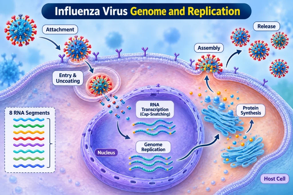
Figure 1: The complete intracellular replication cycle of the influenza A virus, highlighting nuclear entry, cap-snatching transcription, and budding.
1. Anatomy of the Segmented Influenza Genome
Influenza viruses belong to the family Orthomyxoviridae. They are pleomorphic, enveloped virions measuring 80 to 120 nm in diameter. While most standard RNA viruses carry a continuous, unsegmented genome, influenza viruses package their genetic material into discrete, independent negative-sense RNA segments:
- Influenza A and B: Contain 8 distinct RNA segments.
- Influenza C and D: Contain 7 RNA segments.
The 8 RNA Segments of Influenza A:
Segment 1 (PB2) ───> Host Cap Recognition & Polymerase Complex
Segment 2 (PB1) ───> Core Catalytic RNA-Dependent RNA Polymerase
Segment 3 (PA)  ───> Endonuclease for Cap-Snatching
Segment 4 (HA)  ───> Hemagglutinin (Attachment & Membrane Fusion)
Segment 5 (NP)  ───> Nucleoprotein (Packages Genomic vRNA)
Segment 6 (NA)  ───> Neuraminidase (Enzymatic Cleavage & Viral Release)
Segment 7 (M)   ───> M1 Matrix Protein & M2 Proton Ion Channel
Segment 8 (NS)  ───> NS1 (Interferon Antagonist) & NEP/NS2 (Nuclear Export)
The 8 Genomic Segments and Their Encoded Proteins
- Segment 1 (PB2 - Polymerase Basic Protein 2): Recognizes and binds the 5′ methylated caps (m7G) of host cellular pre-mRNAs.
- Segment 2 (PB1 - Polymerase Basic Protein 1): Serves as the central catalytic core of the RNA-dependent RNA polymerase (RdRp), catalyzing phosphodiester bond formation during transcription and replication.
- Segment 3 (PA - Polymerase Acidic Protein): Contains an N-terminal endonuclease domain responsible for cleaving host capped pre-mRNAs during the “cap-snatching” transcription initiation process.
- Segment 4 (HA - Hemagglutinin): The primary surface spike glycoprotein (~80% of surface spikes). Mediates viral attachment to host cell sialic acid receptors and triggers acid-dependent endosomal membrane fusion.
- Segment 5 (NP - Nucleoprotein): Basic structural protein that encapsulates each viral RNA segment, forming helical viral ribonucleoprotein complexes (vRNPs).
- Segment 6 (NA - Neuraminidase): The secondary mushroom-shaped surface glycoprotein (~20% of spikes). A sialidase enzyme that cleaves terminal sialic acid residues to free newly budded virions.
- Segment 7 (M - Matrix Segment): Spliced to yield two vital proteins: M1 (Matrix protein), which forms the structural scaffold beneath the viral envelope, and M2, a homotetrameric proton ion channel embedded in the viral lipid bilayer.
- Segment 8 (NS - Non-Structural Segment): Spliced to produce NS1, a potent immune antagonist that suppresses host type I interferon induction, and NEP/NS2 (Nuclear Export Protein), which coordinates the export of newly synthesized vRNPs from the host nucleus.
2. Why Is Influenza an Error-Prone RNA Virus?
The evolutionary behavior of influenza is governed directly by the biochemistry of its replication enzymes.
Unlike eukaryotic cells and large DNA viruses (such as herpesviruses and poxviruses) that utilize high-fidelity DNA polymerases equipped with 3′→5′ exonuclease proofreading activity, the influenza RNA-dependent RNA polymerase (RdRp) possesses no proofreading or mismatch excision capabilities.
When copying viral RNA, the RdRp introduces nucleotide misincorporations at an exceptionally high frequency: approximately 1 × 10⁻⁴ to 1 × 10⁻⁵ substitutions per nucleotide per replication cycle.
Because the entire influenza A genome is roughly 13,500 nucleotides long, this error rate guarantees that nearly every newly replicated viral genome contains at least one point mutation. Rather than existing as a single static genotype, an influenza population inside an infected lung operates as a dynamic, swarming viral quasispecies—a heterogeneous cloud of closely related genetic variants upon which host immune pressure constantly acts.
3. Step-by-Step Intracellular Replication Cycle
Unlike almost all other negative-sense RNA viruses (which replicate strictly in the host cytoplasm), influenza virus replicates inside the host cell nucleus. This nuclear dependency is required because the virus must exploit host splicing machinery and pre-mRNA capping pathways.
The Influenza Replication Life Cycle:
1. Attachment (HA binds Sialic Acid) ──> 2. Endocytosis & M2 Acidification
3. Nuclear Import of vRNPs ───────────> 4. Cap-Snatching Transcription & Replication
5. Translation in Cytoplasm / ER ─────> 6. Assembly & NA-Mediated Budding Release
Step 1: Attachment to Host Sialic Acid Receptors
Hemagglutinin trimers on the viral surface bind specifically to terminal sialic acid (N-acetylneuraminic acid) residues covalently linked to galactose on host epithelial glycoproteins:
- Human-adapted influenza strains: Preferentially recognize α-2,6-galactose linkages, which predominate on epithelial cells of the human upper respiratory tract (nasopharynx and trachea).
- Avian-adapted influenza strains: Preferentially bind α-2,3-galactose linkages, abundant in the gastrointestinal tracts of waterfowl and the deep alveolar tissues of the human lower respiratory tract.
Step 2: Receptor-Mediated Endocytosis and Uncoating
Following attachment, the virion is internalized via clathrin-dependent endocytosis into an endosome. As the host endosome matures, vacuolar ATPases pump protons into the endosomal lumen, dropping the pH to ~5.0–5.5. This acidification triggers two simultaneous mechanical events:
- HA Conformational Collapse: Acidic pH triggers an irreversible structural rearrangement in HA, exposing a hydrophobic “fusion peptide” that inserts into the endosomal membrane, forcing the viral envelope and endosomal membrane to fuse.
- M2 Ion Channel Opening: The viral M2 protein opens, channeling protons directly from the acidic endosome into the viral interior. This internal acidification breaks electrostatic bonds between the M1 matrix lattice and the encapsulated vRNPs.
Once membrane fusion completes, the disassembled vRNPs spill freely into the host cytoplasm and are rapidly ferried into the nucleus via their nuclear localization signals (NLS) recognized by host importin proteins.
Step 3: Transcription via “Cap-Snatching”
Inside the nucleus, the viral RdRp cannot synthesize 5′ m7G caps or 3′ poly(A) tails independently. Instead, it employs an extraordinary parasitic mechanism termed cap-snatching:
- The PB2 subunit binds directly to the 5′ methylated cap of a newly synthesized host cellular pre-mRNA.
- The PA endonuclease subunit cleaves the host RNA 10 to 14 nucleotides downstream of the cap.
- The hijacked capped host oligonucleotide is then used as a primer by the PB1 subunit to transcribe complementary viral messenger RNA (mRNA).
Step 4: Genome Replication via cRNA Intermediates
To make copies of its negative-sense genome (-vRNA), the viral polymerase uncouples from host-primed transcription and performs unprimed, de novo replication:
Negative-sense vRNA → [RdRp] → Positive-sense cRNA → [RdRp] → Progeny Negative-sense vRNA
The full-length complementary RNA (cRNA) acts as an exact replication intermediate. Newly synthesized -vRNAs are immediately wrapped with nucleoprotein (NP) molecules and capped with RdRp complexes to assemble fresh vRNPs.
Step 5: Protein Synthesis and Glycoprotein Trafficking
Viral mRNAs are exported to the cytoplasm. Host ribosomes translate internal structural and non-structural proteins (M1, NP, NS1, NEP, polymerases).
Simultaneously, the envelope glycoproteins (HA, NA) and the M2 ion channel are translated by membrane-bound ribosomes on the rough endoplasmic reticulum, where they undergo heavy N-linked glycosylation before trafficking through the Golgi apparatus to the apical plasma membrane.
Step 6: Virion Assembly, Budding, and Neuraminidase Release
Progeny vRNPs in the nucleus associate with M1 and NEP/NS2, which engage host exportin-1 (CRM1) to mediate nuclear export into the cytoplasm.
At the plasma membrane, the 8 vRNPs assemble through a highly coordinated segment-specific packaging signal network, ensuring that virtually all viable virions incorporate exactly one copy of each of the 8 distinct segments.
As the virion buds outward through the lipid bilayer, surface hemagglutinin molecules naturally bind back onto the host cell’s own sialic acid residues, tethering newly formed viruses to the dying host cell or causing virions to aggregate with each other.
Here, Neuraminidase (NA) performs its essential function: it enzymatically cleaves terminal sialic acid residues from cell-surface glycoproteins and adjacent virions, freeing the progeny viruses to disperse across the airway mucus layer and infect adjacent epithelial cells.
4. Antigenic Drift: Why Does the Flu Change Every Year?
Antigenic drift is the mechanism responsible for annual influenza epidemics. It represents the continuous, gradual accumulation of point mutations within the genes encoding the globular heads of Hemagglutinin and Neuraminidase.
Antigenic Drift Mechanism:
1. Error-Prone Replication ──> 2. Minor Amino Acid Substitutions in HA/NA
3. Altered Antigenic Epitopes ──> 4. Evasion of Neutralizing Antibodies (Seasonal Flu)
The globular head of HA contains five major hypervariable antigenic loop domains (designated sites A, B, C, D, and E in H3N2; and Sa, Sb, Ca1, Ca2, and Cb in H1N1). Neutralizing antibodies generated by prior infection or vaccination target these exact loops to physically block HA from binding cellular sialic acid.
When single amino acid substitutions emerge within these loops:
- The altered viral protein maintains its ability to bind sialic acid.
- However, pre-existing host antibodies can no longer bind with high affinity.
- The mutant virus enjoys a strong selective growth advantage, evades herd immunity, and rapidly becomes the dominant circulating strain across the population.
This continuous immunological escape explains why seasonal influenza vaccines must be reformulated and readministered virtually every single year.
5. Antigenic Shift: Catastrophic Pandemic Reassortment
While antigenic drift is a gradual drift occurring in both influenza A and B, antigenic shift is an abrupt, catastrophic genetic upheaval unique to Influenza A.
Antigenic shift occurs when an influenza A virus acquires an entirely novel Hemagglutinin segment (with or without a novel Neuraminidase segment) from an animal reservoir, creating an antigenically distinct virus to which the global human population possesses virtually zero pre-existing antibody immunity.
Antigenic Shift via Genetic Reassortment:
[ Human H3N2 Strain ]     [ Avian H1N1 Strain ]
         \                     /
          \                   /
   Co-infection of a "Mixing Vessel" (e.g., Swine)
                   │
                   ▼
  Reassortment of the 8 Segmented RNA Pieces
                   │
                   ▼
       [ Novel Pandemic Reassortant ]
 (e.g., Novel H1N1 with Avian Surface Spikes & Human Core)
The Mechanism of Genetic Reassortment
Because the influenza genome is segmented into 8 independent pieces, if a single host cell (such as a swine respiratory cell, which expresses both human-type α-2,6 and avian-type α-2,3 sialic acid receptors) is simultaneously co-infected by two distinct influenza A strains:
- Both viral genomes disassemble in the host nucleus.
- During packaging, progeny virions can package any mixture of the parental segments.
- With 8 segments, two co-infecting strains can theoretically generate 28 = 256 different reassortant combinations!
Historical Pandemics Driven by Antigenic Shift
- 1918 Spanish Flu (H1N1): Avian-origin strain that adapted directly to human transmission, causing ~50 million deaths worldwide.
- 1957 Asian Flu (H2N2): Reassortment between human H1N1 and an avian H2N2 virus (acquired novel avian HA, NA, and PB1).
- 1968 Hong Kong Flu (H3N2): Reassortment between human H2N2 and an avian H3 virus (acquired novel avian HA and PB1).
- 2009 H1N1 Swine Flu: A complex “quadruple reassortant” virus carrying segments derived from North American classical swine, North American avian, human seasonal H3N2, and Eurasian swine lineages.
6. Antigenic Drift vs. Antigenic Shift: Core Differences
| Feature | Antigenic Drift | Antigenic Shift |
|---|---|---|
| Nature of Genetic Change | Minor, gradual point mutations (substitutions) | Major, abrupt structural replacement of entire segments |
| Biochemical Mechanism | RdRp replication errors without proofreading | Genetic reassortment during dual viral co-infection |
| Genomic Substrate | Nucleotide sequence within individual genes | Exchange of whole RNA segment(s) |
| Influenza Types Affected | Influenza A and Influenza B | Strictly Influenza A (due to animal reservoirs) |
| Frequency of Occurrence | Continuous (annual / seasonal) | Rare and unpredictable (decadal intervals) |
| Epidemiological Outcome | Seasonal epidemics; localized outbreaks | Global pandemics with high morbidity |
| Immune Protection | Partial cross-protection from past infections | Zero pre-existing herd immunity in humans |
| Vaccine Requirement | Annual updates to seasonal vaccine formulation | Emergency development of a novel pandemic vaccine |
7. Decoding Subtype Nomenclature: Why H1N1, H3N2, and H5N1?
The classification system for influenza A viruses is defined by the antigenic properties of its two primary surface glycoproteins:
- Hemagglutinin (H): 18 recognized subtypes (H1 through H18).
- Neuraminidase (N): 11 recognized subtypes (N1 through N11).
Subtypes H1 to H16 and N1 to N9 circulate widely in wild aquatic waterfowl—the natural evolutionary reservoir for all influenza A viruses. Subtypes H17N10 and H18N11 have been identified exclusively in bats.
Only three hemagglutinin subtypes (H1, H2, and H3) and two neuraminidase subtypes (N1 and N2) have established sustained, endemic transmission cycles in the human population. Highly pathogenic avian influenza strains, such as H5N1 and H7N9, can cross the species barrier directly from poultry to humans, causing fatal pneumonia; however, because they have not yet acquired mutations allowing efficient human-to-human transmission via airborne droplets, they remain zoonotic spillover threats rather than global pandemics.
8. Global Surveillance and Seasonal Vaccine Formulation
Because influenza undergoes relentless antigenic drift, seasonal vaccines require constant recalibration.
The World Health Organization Global Influenza Surveillance and Response System (GISRS) monitors circulating viruses across 150+ national influenza centers globally.
Twice each year (in February for the Northern Hemisphere and in September for the Southern Hemisphere), international advisory committees analyze thousands of clinical isolates using:
- Hemagglutination Inhibition (HI) Assays: Measuring antibody neutralization titers against emerging strains.
- Next-Generation Whole-Genome Sequencing: Tracking phylogenetic clade emergence and amino acid shifts across the HA and NA globular heads.
- Antigenic Cartography: Mathematical modeling visualizing antigenic distance between circulating variants.
Based on these data, the WHO recommends the composition for trivalent or quadrivalent seasonal influenza vaccines (typically incorporating two Influenza A strains—H1N1 and H3N2—and one or two Influenza B lineages), giving manufacturers six months to produce hundreds of millions of doses in embryonated chicken eggs or cell cultures before the winter flu season arrives.
Conclusion
The influenza virus is a masterclass in microbial evolution. Its single-stranded negative-sense RNA genome, error-prone polymerase, and segmented structure provide an evolutionary architecture engineered for survival and escape.
Through antigenic drift, the virus constantly polishes its surface antigens, outrunning our adaptive immune memory year after year. Through antigenic shift, it periodically draws a completely new genetic hand from animal reservoirs, resetting human herd immunity to zero and testing the limits of global public health readiness.
Understanding the molecular mechanisms governing influenza genome replication and antigenic variation is essential not only for medical virologists but for anyone interested in infectious disease epidemiology. As structural vaccinology advances toward “universal” influenza vaccines targeting the conserved stalk of the hemagglutinin molecule, molecular insights into this adaptable pathogen bring us closer to breaking the cycle of seasonal epidemics and pandemic threats for good.