- 1 What Are Cytopathic Effects (CPE)?
- 2 Why Are CPE Important in Virology?
- 3 Major Types of Cytopathic Effects Observed in Cell Culture
- 4 Common Viral Infection Patterns in Cell Culture
- 5 How to Identify CPE Under a Microscope
- 6 Why Some Viruses Do Not Produce Visible CPE
- 7 Can CPE Alone Be Used to Identify a Specific Virus?
- 8 Key Takeaways
Imagine looking at a healthy cell culture under a microscope. The cells are attached to the surface, have a relatively uniform appearance, and form a continuous monolayer. Now imagine examining the same culture after viral exposure. Some cells have become rounded, others appear enlarged, clusters are forming, and parts of the monolayer are beginning to detach.
These visible changes may be signs of a viral infection. In virology, they are known as cytopathic effects (CPE). CPE are among the most important visual clues used by researchers and diagnostic laboratories to monitor viral growth in cell culture. Different viruses can produce different patterns of cellular damage, and learning to recognize these patterns helps researchers understand what may be happening inside an infected culture.
However, an important principle must be kept in mind: not every damaged cell is infected by a virus, and not every virus produces visible CPE. This is why careful observation and confirmatory testing are essential.
In this guide, we will explore the major types of cytopathic effects, how to recognize them under a microscope, examples associated with common viruses, and how to distinguish genuine viral infection patterns from other cell culture problems.
What Are Cytopathic Effects (CPE)?
Cytopathic effects are structural or morphological changes that occur in host cells as a result of viral infection. When a virus enters a susceptible cell, it uses the host’s cellular machinery to produce viral components and, in many cases, new viral particles.
During this process, normal cellular functions may become disrupted. The infected cell can change its shape, lose attachment to the culture surface, fuse with neighboring cells, or eventually die. These changes can be observed using a light microscope and are collectively described as cytopathic effects.
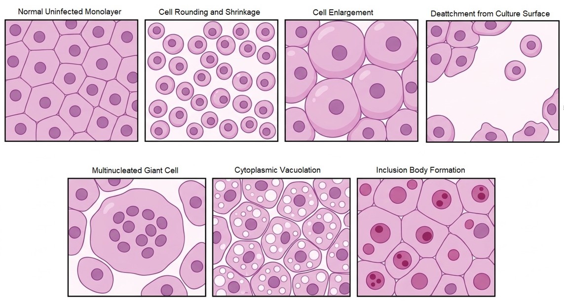
Common CPE include:
- Cell rounding and shrinkage
- Cell enlargement (cytomegaly)
- Cell detachment from the culture surface
- Formation of multinucleated giant cells (syncytia)
- Cytoplasmic vacuolation
- Inclusion body formation (intranuclear or intracytoplasmic)
- Cell lysis and destruction of the monolayer
The appearance and severity of CPE depend on several factors, including the virus, host cell type, viral strain, infection conditions, and stage of infection. Importantly, some viruses produce obvious cellular damage, while others cause very subtle changes or no visible CPE at all.
Why Are CPE Important in Virology?
CPE provide a visual indication that something may be happening in a cell culture following viral exposure. Traditionally, researchers and diagnostic laboratories have used microscopic examination of cell monolayers to monitor viral growth. Changes in cellular morphology can provide an early indication of infection and help guide further investigation.
CPE are useful for several reasons:
- Monitoring viral growth: Tracking the progression of infection over hours or days.
- Recognizing possible viral infection patterns: Associating specific morphological phenotypes with candidate viral families.
- Evaluating cell damage: Measuring the cytopathic potency and kinetics of viral isolates.
- Supporting viral identification: Narrowing down differentials before deploying targeted molecular or immunological assays.
Major Types of Cytopathic Effects Observed in Cell Culture
Let us look at the most common CPE patterns and what they look like under the microscope.
1. Cell Rounding
Cell rounding is one of the most commonly observed early morphological changes during viral infection. Healthy adherent cells often maintain their characteristic shape (such as elongated fibroblasts or polygonal epithelial sheets) and remain firmly attached to the culture surface.
Following viral infection, affected cells may lose their normal shape and become rounded. They may also appear more refractile, meaning they reflect light differently and become brighter and easier to distinguish from surrounding cells. As cellular damage progresses, rounded cells may detach from the surface.
What to look for:
- Loss of normal cell shape
- Rounded or spherical appearance
- Increased refractility under phase-contrast or brightfield microscopy
- Reduced attachment to the culture surface
- Gaps appearing in the monolayer
Cell rounding can occur with several viruses, including herpes simplex virus (HSV) and enteroviruses, although the exact appearance depends on the virus and cell line.
2. Syncytium Formation (Multinucleated Giant Cells)
Syncytium formation is one of the most recognizable cytopathic effects. Some viruses produce fusion surface glycoproteins that promote the fusion of infected cells with neighboring uninfected cells. Instead of remaining separate, multiple cells merge and form a large, continuous structure containing several nuclei within a shared cytoplasm. This structure is called a syncytium (plural: syncytia).
Under the microscope, syncytia may appear as unusually large cells containing multiple nuclei clustered together.
What to look for:
- Large fused cellular structures
- Multiple nuclei within a shared cytoplasm
- Irregular cell boundaries
- Groups of cells appearing merged together
Viruses commonly associated with syncytium formation include:
- Respiratory syncytial virus (RSV)
- Measles virus
- Certain parainfluenza viruses
- Human immunodeficiency virus (HIV) in CD4+ cell lines
3. Inclusion Body Formation
Inclusion bodies are abnormal, discrete structures that appear inside the nucleus or cytoplasm of infected cells. They can represent accumulations of assembled viral components, crystalline arrays of viral particles, altered host-cell structures, or combinations of both. Their appearance and sub-cellular location provide essential diagnostic clues.
There are two broad categories:
A. Intranuclear Inclusion Bodies
These are found inside the nucleus of the infected cell. Herpes simplex virus and adenoviruses are well-known examples associated with characteristic intranuclear inclusions (e.g., Cowdry type A inclusions in HSV).
B. Intracytoplasmic Inclusion Bodies
These appear within the cytoplasm. Rabies virus, for example, is classically associated with cytoplasmic Negri bodies in infected neuronal tissue. Similarly, poxviruses produce prominent eosinophilic cytoplasmic inclusions (Guarnieri bodies).
What to look for:
- Distinct, stained or refractile structures inside the nucleus or cytoplasm
- Changes in nuclear appearance and altered chromatin distribution (e.g., margination of chromatin)
- Clear halo surrounding an inclusion body
Note: Inclusion bodies are not always easily visible in living, unstained cultures. Fixation and histological staining (such as Giemsa, hematoxylin & eosin, or Papanicolaou stains) are typically required to identify them clearly.
4. Cell Enlargement (Cytomegaly)
Some viral infections cause cells to become unusually large. This phenomenon is known as cytomegaly. Instead of becoming smaller or rounded, affected cells increase markedly in volume and develop abnormal nuclear or cytoplasmic features.
Cytomegalovirus (CMV), as its name suggests, is the prototypical example associated with dramatically enlarged infected cells, often displaying prominent intranuclear inclusions resembling an “owl’s eye.”
What to look for:
- Marked enlargement of cellular diameter
- Abnormally large nuclei
- Characteristic nuclear chromatin alterations
- Accompanying inclusion bodies
5. Cytoplasmic Vacuolation
Vacuolation refers to the appearance of clear, bubble-like spaces (vacuoles) within the cytoplasm. During certain viral infections, disruption of membrane trafficking, endoplasmic reticulum stress, and altered cellular lipid/water metabolism contribute to the rapid accumulation of cytoplasmic vacuoles. Under the microscope, affected cells may look granular, bubbly, or foamy.
What to look for:
- Clear, distinct spaces within the cytoplasm
- Granular or foamy cellular appearance
- Disruption of normal intracellular architecture
- Progressive cellular degeneration
Important consideration: Vacuolation is not specific to viral infection. It can also occur as a consequence of chemical toxicity, pH fluctuations, media nutrient exhaustion, or cellular stress.
6. Cell Detachment and Lysis
In strongly cytolytic viral infections, cellular damage progresses until affected cells lose their attachment to the culture vessel surface. Eventually, cells undergo complete lysis, releasing progeny virions and intracellular contents into the surrounding culture medium. This results in progressive thinning, visible gaps, and complete destruction of the cell monolayer.
What to look for:
- Increasing numbers of detached, floating cells
- Floating cellular debris and granular fragments
- Loss of monolayer integrity
- Large empty areas in the culture vessel
- Extensive cellular destruction
This type of CPE is particularly noticeable in enterovirus (e.g., poliovirus, coxsackievirus) and adenovirus infections.
Common Viral Infection Patterns in Cell Culture
Different viruses produce recognizable CPE patterns in specific cell lines. However, the same virus can look different depending on the host cell substrate used, while unrelated viruses can sometimes produce similar appearances.
The following table provides classic examples:
| Virus | Commonly Reported CPE Pattern |
|---|---|
| Herpes simplex virus (HSV) | Cell rounding, enlargement, ballooning, and rapid degeneration |
| Adenovirus | Rounded cells aggregating into grape-like clusters or lacy monolayers |
| Respiratory syncytial virus (RSV) | Extensive syncytium formation in susceptible epithelial lines |
| Measles virus | Multinucleated giant-cell (syncytial) formation |
| Cytomegalovirus (CMV) | Marked cytomegaly (enlarged cells) with classic “owl’s eye” inclusions |
| Enteroviruses | Rapid cell rounding, refractility, shrinkage, and complete monolayer lysis |
| Parainfluenza viruses | Cell rounding and, in certain cell lines, syncytia |
| Influenza virus | Granular degeneration, cell detachment, and variable cellular changes |
These are general patterns rather than absolute identification criteria. The observed CPE must always be interpreted alongside the cell line origin, specimen passage history, and confirmatory laboratory assays.
How to Identify CPE Under a Microscope
Recognizing CPE is not simply about looking for damaged cells. It involves systematically comparing the appearance of a culture over time and identifying changes that diverge from normal cellular growth.
Here are key principles for microscopic examination:
1. Understand the Appearance of Healthy Cells
Before attempting to identify CPE, become thoroughly familiar with the normal morphology of the specific cell line being examined. Healthy cells typically show:
- Consistent cell shape and uniform orientation
- Appropriate, firm attachment to the culture surface
- Relatively uniform distribution across the vessel
- An intact, confluent monolayer (where applicable)
- Absence of excessive floating cellular debris
Remember: different cell lines naturally display different morphologies. For instance, epithelial-like (polygonal) and fibroblast-like (spindle-shaped) cells look entirely different in their uninfected state.
2. Always Compare Infected and Uninfected Controls
One of the most critical steps in CPE interpretation is comparing the inoculated test culture with an uninoculated negative control maintained in parallel under identical media, incubation, and temperature conditions. This verifies whether observed morphological alterations are genuinely linked to viral replication or represent media toxicity, temperature stress, or aging cell monolayers.
3. Look for Combinations of Morphological Changes
Pay close attention to:
- Rounding and shrinkage
- Cell enlargement and swelling
- Clustering or plaque formation
- Cytoplasmic vacuolation
- Cell fusion and syncytia
- Detachment and monolayer destruction
A constellation of multiple cellular alterations provides substantially stronger evidence than a single non-specific abnormal feature.
4. Observe the Distribution of Cellular Damage
Viral infection often begins as localized foci of cellular change, which progressively expand across the monolayer as progeny virions spread to adjacent cells. In contrast, non-specific toxicities (e.g., pH shifts or toxic reagents) typically cause generalized, uniform damage across the entire culture vessel simultaneously.
5. Record Changes Systematically Over Time
CPE develops dynamically. A single snapshot examination may yield incomplete or ambiguous information. Maintaining standardized daily observation logs and capturing photomicrographs at 24, 48, and 72 hours post-inoculation allows researchers to trace the kinetics of cellular alterations accurately.
Why Some Viruses Do Not Produce Visible CPE
A frequent misconception in diagnostic virology is that every viral infection inevitably causes overt cellular destruction. This is not the case:
- Some viruses replicate efficiently in susceptible cells without inducing noticeable morphological alterations (non-cytopathic viruses).
- Others cause very subtle or delayed changes that evade routine brightfield microscopy.
- Certain persistent or latent infections maintain host-cell viability while continuously shedding low levels of viral particles.
The absence of visible CPE does not rule out active viral infection. In such instances, supplementary diagnostic and analytical methodologies are indispensable:
- Reverse Transcription Polymerase Chain Reaction (RT-PCR) / qPCR: Detection of viral nucleic acids.
- Immunofluorescence Assays (IFA): Direct or indirect detection of viral antigens within intact cells.
- Hemadsorption: Adsorption of red blood cells onto the surface of infected cells expressing viral hemagglutinin (e.g., influenza, parainfluenza).
- Enzyme-Linked Immunosorbent Assays (ELISA): Detection of viral proteins secreted into the culture supernatant.
Can CPE Alone Be Used to Identify a Specific Virus?
While CPE provides valuable diagnostic clues and guides laboratory workflow, morphology alone is rarely sufficient to identify a virus definitively. Multiple unrelated viruses can produce indistinguishable cell rounding or syncytia, and viral CPE can vary considerably across different cell passage levels and host backgrounds.
Reliable viral identification requires integrating several parameters:
- The host cell substrate (species, tissue origin, primary vs. continuous line).
- The exact morphology, timeline, and spatial pattern of the CPE.
- Clinical history, specimen source, and patient symptoms.
- Confirmatory molecular or immunological testing (PCR, antigen testing, or sequencing).
Modern virology relies on the synergy of cell culture observation and molecular precision.
Key Takeaways
Cytopathic effects are far more than damaged cells under a microscope—they represent a direct visual window into the pathogenic mechanisms operating between viruses and host cells. From cell rounding and detachment to syncytium formation and diagnostic inclusion bodies, each pattern reveals key aspects of viral biology.
For students, researchers, and diagnostic scientists, mastering CPE recognition is foundational to virology. By combining careful microscopy with parallel uninfected controls, contamination screening, and confirmatory molecular assays, laboratories achieve precise and reliable viral diagnosis.