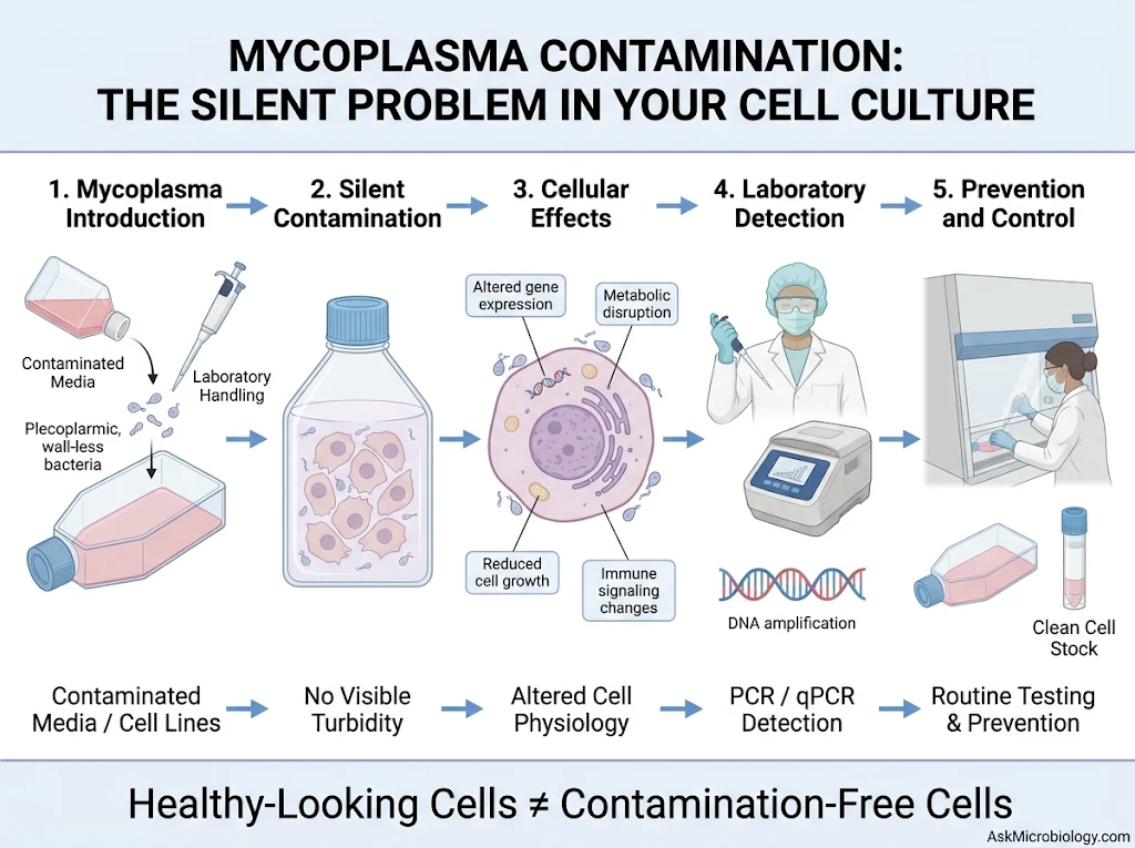
- 1 What Is Mycoplasma and Why Is It Different from Other Bacteria?
- 2 Why Is Mycoplasma Contamination Called a Silent Problem?
- 3 How Does Mycoplasma Enter a Cell Culture Laboratory?
- 4 How Does Mycoplasma Affect Your Cells?
- 5 How Can You Detect Mycoplasma Contamination?
- 6 What Should You Do If Your Cell Culture Tests Positive?
- 7 How Can You Prevent Mycoplasma Contamination?
- 8 Why Is Mycoplasma Testing Important Before RNA-seq, qPCR, and Cell-Based Experiments?
- 9 Conclusion
Imagine spending several weeks growing cells for an important experiment. Your cells look healthy under the microscope, the culture medium is clear, and everything appears to be working perfectly. You proceed with RNA extraction, qPCR, protein analysis, or even sequencing. But there is one problem: your cell culture is contaminated. Not with an obvious bacterial or fungal contaminant, but with something much harder to notice: Mycoplasma. Unlike conventional bacterial contamination, mycoplasma can remain hidden in cell cultures without producing obvious changes in the appearance of the medium. Your cells may continue growing, but their metabolism, gene expression, and biological behavior can gradually change. This is why mycoplasma contamination is often called the silent enemy of cell culture laboratories. For researchers working with mammalian cell lines, primary cells, stem cells, or immune cells, this contamination can become a serious problem. It can compromise experimental results, affect cell viability, and sometimes invalidate months of research. So, what makes mycoplasma so difficult to detect? How does it enter cell cultures? And most importantly, how can laboratories prevent it?
Let’s explore the microbiology behind this common laboratory problem.
What Is Mycoplasma and Why Is It Different from Other Bacteria?
Mycoplasmas are among the smallest self-replicating prokaryotic microorganisms known. They belong to the class Mollicutes and are characterized by one particularly unusual feature: they completely lack a conventional cell wall. Most bacteria possess a peptidoglycan cell wall that provides structural support and maintains cellular shape. Mycoplasmas do not have this structure. Instead, their cell membrane contains sterols that help maintain membrane stability. This unique biology gives them several characteristics that make them difficult to control in cell culture laboratories.
They are extremely small, generally around 0.2–0.8 µm.
They can pass through some filters designed to remove conventional bacteria.
They are naturally resistant to antibiotics that target cell wall synthesis, such as penicillin.
Their flexible cell membranes allow considerable variation in shape.
They can survive and multiply in association with cultured cells.
Some of the most frequently encountered cell culture contaminants include:
- Mycoplasma orale
- Mycoplasma hyorhinis
- Mycoplasma arginini
- Mycoplasma fermentans
- Mycoplasma hominis
- Mycoplasma salivarium
- Mycoplasma pirum
- Acholeplasma laidlawii
Although these organisms belong to different species and genera, they share the ability to interfere with normal cell culture conditions.
Why Is Mycoplasma Contamination Called a Silent Problem?
This is probably the most frustrating characteristic of mycoplasma contamination. When ordinary bacteria contaminate a cell culture, the signs are often relatively easy to recognize. The medium may become cloudy, its pH may change, and bacterial growth may become visible under the microscope. Mycoplasma behaves differently.
In many contaminated cultures:
- The culture medium remains clear.
- No obvious turbidity develops.
- Cells may continue growing.
- The normal cell morphology may appear unchanged.
- Routine microscopic examination may fail to reveal contamination.
As a result, researchers can unknowingly maintain contaminated cells for multiple passages. The real problem is that normal-looking cells are not necessarily biologically healthy cells. A culture may appear perfectly acceptable while mycoplasma is already influencing cellular metabolism, proliferation, gene expression, and immune responses. This makes visual inspection alone insufficient for confirming that a cell culture is contamination-free.
How Does Mycoplasma Enter a Cell Culture Laboratory?
Mycoplasma contamination does not always originate from poor laboratory technique. It can enter through several routes, including contaminated cultures, reagents, equipment, and handling procedures. Understanding these sources is the first step toward preventing contamination.
Contaminated Cell Lines
One of the most important sources is an already contaminated cell culture obtained from another laboratory or a commercial supplier. When researchers introduce a new cell line without testing it, they may unknowingly introduce mycoplasma into an otherwise clean laboratory environment. If the contaminated culture is handled alongside other cell lines, cross-contamination can occur.
Laboratory Personnel
Human handling is another possible source. Contamination can spread through improperly handled pipettes, shared equipment, contaminated gloves, and the transfer of materials between clean and infected cultures. For example, using the same pipette or reagent bottle for multiple cultures without appropriate precautions can introduce contaminants into previously clean samples.
Culture Media and Reagents
Media, serum, supplements, and other culture reagents can potentially introduce mycoplasma if their quality is not adequately controlled. Although reputable commercial suppliers generally apply quality-control procedures, researchers should still pay attention to product certification and laboratory storage and handling practices.
Laboratory Equipment
Incubators, biosafety cabinets, water baths, and other shared equipment can become involved in contamination events. Improper cleaning and the handling of contaminated materials can contribute to the spread of microorganisms between cultures. An important point is that a biosafety cabinet does not automatically guarantee contamination-free work. Proper cleaning, maintenance, and aseptic technique remain essential.
How Does Mycoplasma Affect Your Cells?
Mycoplasma contamination is not simply an unwanted microorganism growing alongside your cells. It can actively change how those cells behave. This is particularly concerning when researchers are studying gene expression, cellular metabolism, immune signaling, or drug responses.
Let’s look at some important consequences.
Changes in Cell Growth and Proliferation
Mycoplasmas compete with host cells for nutrients and essential metabolic components. Some species can consume important nutrients, including amino acids such as arginine. This competition may interfere with normal cell growth and alter proliferation rates. Consequently, researchers may observe unexpected differences in cell numbers or growth behavior between experimental groups.
Altered Gene Expression
One of the most serious consequences is the disruption of normal gene expression. Mycoplasma infection can influence cellular signaling pathways, inflammatory responses, and transcriptional activity. This means that gene expression changes observed during an experiment may not necessarily result from the treatment being investigated. For example, imagine studying the expression of an immune-related gene after stimulating T cells. If the cells are contaminated with mycoplasma, the infection itself may influence immune signaling and alter the expression of the target gene. The resulting qPCR data could therefore be misleading.
Changes in Cellular Metabolism
Mycoplasmas can interfere with several metabolic pathways, including amino acid, nucleotide, and energy metabolism. These changes may affect ATP production, cellular stress responses, and the availability of important metabolic intermediates. For researchers performing metabolomics or studying cellular energy regulation, this contamination can become particularly problematic.
Effects on Chromosomes and DNA
Some mycoplasma species produce enzymes and other factors that can affect host-cell DNA and cellular signaling. Prolonged contamination has been associated with chromosomal abnormalities and changes in DNA synthesis. This can complicate experiments involving genomic analysis, cell differentiation, and long-term cell culture.
Reduced Transfection Efficiency
Mycoplasma contamination can also interfere with transfection experiments. Researchers may observe inconsistent transfection efficiency, altered expression of introduced genes, or unexpected cellular responses following transfection. This is particularly important when working with plasmids, CRISPR-based systems, viral vectors, or other genetic engineering approaches. For laboratories studying gene editing and cellular therapies, maintaining mycoplasma-free cultures is essential for reliable experimental interpretation.
How Can You Detect Mycoplasma Contamination?
Since mycoplasma is difficult to identify through ordinary microscopic examination, specialized detection methods are required. Several techniques are commonly used in cell culture laboratories.
PCR-Based Detection
PCR is one of the most widely used approaches for identifying mycoplasma contamination. The method detects specific DNA sequences associated with mycoplasma organisms.
A typical PCR-based assay involves:
- Collecting an appropriate culture supernatant or cell-associated sample.
- Extracting microbial DNA or preparing the sample according to the assay protocol.
- Amplifying conserved mycoplasma-associated genetic targets.
- Analyzing the amplification products through gel electrophoresis or an appropriate detection platform.
- Comparing the results with positive and negative controls.
A positive amplification signal suggests the presence of mycoplasma DNA. However, PCR can also detect DNA from nonviable organisms. Therefore, a positive result does not automatically demonstrate that viable mycoplasma is actively multiplying in the culture.
Quantitative PCR (qPCR)
Real-time PCR provides a sensitive approach for detecting mycoplasma DNA. Unlike conventional endpoint PCR, qPCR monitors amplification during the reaction using fluorescent signals. This allows laboratories to detect contamination without necessarily requiring post-PCR gel electrophoresis. Some commercial assays are designed to detect multiple mycoplasma species using conserved genomic targets. However, assay coverage depends on the primers and probes used, so laboratories should check the manufacturer’s validation information.
DNA Staining Methods
Fluorescent DNA-binding dyes, such as Hoechst 33258 or DAPI, can be used to visualize mycoplasma-associated DNA. Under fluorescence microscopy, contamination may appear as small fluorescent particles or filamentous structures around the cell surface. These methods can provide a useful visual indication of contamination, although interpretation requires experience. They may also produce misleading results because extracellular DNA and other microorganisms can generate fluorescence.
Mycoplasma Culture Methods
Specialized microbiological culture methods can also be used to detect viable mycoplasma organisms. Unlike ordinary bacterial culture, these methods require specialized media and incubation conditions. They can take considerably longer than molecular detection methods, sometimes requiring several weeks. However, culture-based testing can provide evidence of viable organisms and may help identify contaminants that are not detected by a particular molecular assay.
Which Detection Method Is Best?
For routine cell culture quality control, validated PCR-based methods are widely used because they provide a practical balance between sensitivity, speed, and reliability. Where necessary, laboratories can combine molecular and culture-based approaches for confirmation.
| Method | Main advantage | Main limitation |
|---|---|---|
| Conventional PCR | Relatively simple and accessible | Requires careful interpretation of amplification results |
| qPCR | Sensitive and rapid detection | Depends on assay coverage and validation |
| DNA staining | Provides visual evidence | Less specific than molecular methods |
| Culture method | Can demonstrate viable organisms | Slow and technically demanding |
What Should You Do If Your Cell Culture Tests Positive?
Discovering mycoplasma contamination can be frustrating, especially when you have spent weeks or months preparing cells for an important experiment. However, the first step should be to prevent the contamination from spreading. Here is a sensible laboratory response.
Step 1: Isolate the suspected culture
Immediately separate the potentially contaminated culture from other cell lines. Avoid sharing media, pipettes, and reagents between contaminated and clean cultures.
Step 2: Confirm the result
Repeat testing using an appropriate validated assay and suitable controls. If necessary, use an alternative detection method to investigate the result.
Step 3: Evaluate the value of the cell line
If the cell line is readily available from an authenticated, mycoplasma-free source, discarding the contaminated culture and restarting from a clean stock is often the safest option. For valuable or difficult-to-replace cell lines, specialized mycoplasma elimination treatments may be considered.
Step 4: Decontaminate the working environment
Clean and disinfect the relevant biosafety cabinet, incubator, equipment, and work surfaces using procedures appropriate for the suspected contaminant. Follow institutional biosafety and equipment-manufacturer recommendations.
Step 5: Retest before using the cells
Never assume that treatment has successfully eliminated contamination. The culture should be tested again after treatment and recovery, according to a validated testing schedule. A negative result should be confirmed at appropriate follow-up intervals before the cells return to experimental use. One important warning: simply adding antibiotics to contaminated cultures is not a reliable solution. Many conventional cell culture antibiotics do not eliminate mycoplasma.
How Can You Prevent Mycoplasma Contamination?
Prevention is considerably easier than trying to rescue an infected cell line. A good laboratory routine can significantly reduce the risk of contamination.
Here are some practical measures:
- Test newly received cell lines before introducing them into established cultures.
- Maintain separate handling procedures for different cell lines.
- Use sterile, properly maintained pipettes and culture materials.
- Avoid sharing reagent bottles between cultures.
- Regularly clean biosafety cabinets and incubators.
- Use appropriate personal protective equipment.
- Maintain good aseptic technique during cell passaging and media replacement.
- Purchase media, sera, and supplements from reliable suppliers.
- Maintain frozen stocks of authenticated, mycoplasma-free cells.
- Perform routine mycoplasma testing rather than relying on visual inspection.
- Avoid using antibiotics as a substitute for proper contamination control.
Routine testing is particularly important for laboratories working with primary cells, stem cells, immune cells, and long-term cultures.
Why Is Mycoplasma Testing Important Before RNA-seq, qPCR, and Cell-Based Experiments?
This is an area that deserves particular attention because contamination can influence the interpretation of molecular biology experiments. Imagine that you are comparing gene expression between treated and untreated cells. You isolate RNA, prepare cDNA, perform qPCR, and observe a significant difference in your target gene. You may conclude that your treatment has successfully changed gene expression. But what if one experimental group was contaminated with mycoplasma? The observed difference could partly reflect infection-induced changes in cellular signaling rather than the biological effect of your treatment.
The same concern applies to several other techniques:
- RNA-seq: Mycoplasma can alter host gene expression and complicate transcriptomic interpretation.
- qPCR: Infection-related changes can influence target gene expression and reference-gene stability.
- Western blotting: Altered cellular signaling may change protein abundance.
- Transfection: Contamination can interfere with cellular responses and transfection efficiency.
- CRISPR experiments: Changes in cell physiology can affect editing outcomes and experimental reproducibility.
- Cell viability assays: Altered metabolism may influence assay signals independently of actual cell numbers.
- ATAC-seq: Mycoplasma-derived DNA can interfere with sequencing-based analyses of chromatin accessibility.
This is why testing cells before important experiments is not just a quality-control formality. It is part of experimental validation.
Conclusion
Mycoplasma contamination is one of the most challenging problems in cell culture because it can remain hidden while gradually affecting cellular biology. Unlike conventional bacterial contamination, it may not produce cloudy media, obvious morphological changes, or rapid cell death. Instead, it can interfere with metabolism, proliferation, gene expression, immune signaling, and experimental reproducibility. The good news is that mycoplasma contamination can be controlled through proper laboratory practices, routine molecular testing, appropriate isolation procedures, and reliable cell banking. For every researcher working with cultured cells, one principle is worth remembering:
Healthy-looking cells are not necessarily contamination-free cells.
A culture may look perfect under the microscope, but without appropriate testing, its biological reliability cannot be guaranteed. Sometimes, the smallest organisms create the biggest problems in the laboratory.